Image Analysis
From cell imaging to high-throughput counting and tracking
Manual identification, counting and tracking of cells, while useful as a quick tool for smaller projects, quickly becomes difficult or outright impossible as the backlog of images increases to the hundreds or thousands.
This process is also prone to errors and fluctuations, even when performed by trained individuals. Automation is important.
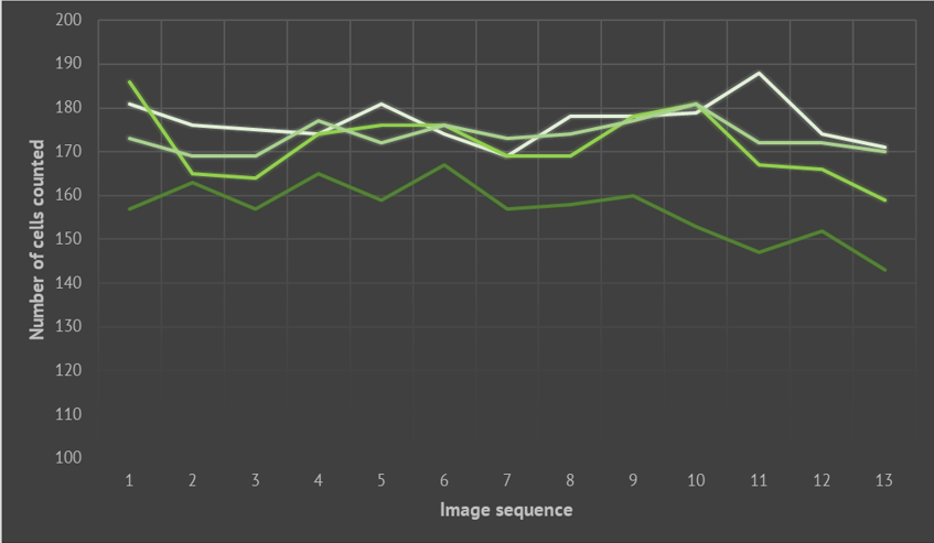
Manual cell counting comparison. Lines represent counts performed by different lab members.
Existing image analysis tools
Many open source solutions such as Icy and CellProfiler exist.
These software include a vast array of modules, plugins and pipelines to detect cells and analyze biological images.


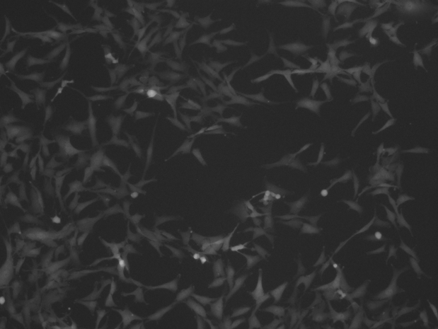
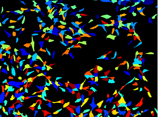
Example of object detection as performed by CellProfiler.
Our current work
Our current work focuses on these aspects of image analysis:
Incorporation of programming languages into biological sciences, allowing for faster data analysis.
Tracking cells across multiple images, allowing reconstruction of cell destiny and colony family trees.
Overcoming challenges in cell segmentation software: correctly detecting cells close to each other and cell colonies.
Creation of an integrated and automated pipeline, from long-time image acquisition all the way to data visualization.
If you would like to propose a collaboration, contact us .