
Health | Research shows that, in newborn rats, the virus causes changes in developmental milestones and in the blood-brain barrier that protects the nervous system
*By Alexandre Briozo Gomes Filho
*First published July 14. 2022
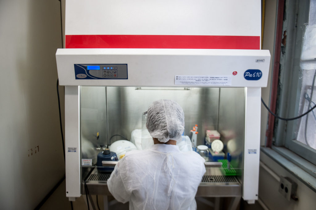
*Photo: Flávio Dutra/JU archive April 9, 2020
On November 15, 2015, the Ministry of Health recognized the Zika virus outbreak as a National Health Emergency, with the largest concentration occurred in the Northeast – mainly in poor and peripheral neighborhoods, places without basic sanitation that facilitate the proliferation of the virus vector, the Aedes aegypti mosquito. The period coincided with the significant increase in cases of fetal microcephaly (brain malformation in the prenatal period), and for some time this coincidence was not officially associated with the occurrences of microcephaly, despite health surveillance in the region having signaled causality between the virus and the condition. In 2015 alone, 2,782 cases and 40 deaths were recorded from microcephaly. Later, with the efforts of both local and foreign scientists, causality was confirmed through laboratory research.
Despite the containment of the Zika virus outbreak and the reduction in cases of microcephaly, the research on the topic has not ceased, on the contrary: it has been moving towards new answers. This is the case of a study by the Federal University of Rio Grande do Sul (UFRGS), published in the journal Brain, Behavior and Immunity, which observed that the virus poses after-effects which go beyond microcephaly, not only in humans but also in rats. The researchers detected that the presence of the virus in the rats’ bodies caused changes in the blood-brain barrier (BBB), a structure that prevents the passage of substances from the blood to the central nervous system. This change is associated with the oxidative stress noted in these animals due to contamination.
Important changes for their survival were also noted in the newborns’ developmental milestones. The clinical identification of these effects in rats – despite having previously been identified in humans – is important so that the animal model can serve as a source of study for this pathology and support future treatments. The study was carried out by researchers from the Institute of Basic Health Sciences (ICBS) at UFRGS, the Federal University of Pelotas, and the Institute of Cardiology.
From methods to results
“We proposed to inoculate the virus in pregnant rats on the ninth day of pregnancy, a critical period in the development of the rodents’ nervous system,” explains neuroscientist and professor at ICBS/UFRGS Mrs. Lenir Orlandi, leader of the research. “The rats did not develop diseases and no mortality was observed, unlike what would be occasional for this type of work,” says Professor Orlandi.
In newborn rats inoculated with the virus, developmental delays were observed after 22 days of the rodents’ neonatal period, even though the newborns had been born with unchanged brain size. These delays range from ear release the— which first develops stuck together and then opens with increased hearing expression — to nest and the mother localization through smell. After this period, the animals were euthanized to assess whether the number of important cells for structures – such as the hippocampus, for example – had been reduced, but no changes were found.
However, when measuring the proteins of the blood-brain barrier that protects the nervous system, the group of researchers noticed a decrease in several of these substances, showing that the brains of those rodents were possibly more susceptible to the presence of the virus. This decrease in proteins is associated with the oxidative stress noted in newborns, indicating the disordered presence of free radicals (a byproduct of chemical reactions) in the nervous system, which can cause damage to cells if not neutralized by antioxidants. Oxidative stress occurs when the body’s antioxidant levels are not high enough to combat the harmful effects of free radicals.
“We also evaluated the mothers’ placenta and the tissue of the fetuses, and we saw that the virus was in the fetal material, it was in the placenta,” explains the neuroscientist. She also explains that the changes in the nervous system are justified by the greater attraction that the virus has for the cells of this system.
“The Zika virus is attracted to precursor neuronal cells present in the prenatal period – precisely the cells that originate all the other ones of our nervous system.”
Lenir Orlandi
Similar effects of congenital Zika virus syndrome have already been studied in humans, but finding they may also be present in rats makes it possible to reproduce, in rodents, what happens in humans and allows researchers to more properly and deeply investigate such after-effects and maybe find new therapeutic strategies
SARS-CoV-2 on the bus and Zika virus at CREAL
When the group’s article was accepted for submission by Brain, Behavior, and Immunity, a high-impact journal in the scientific scene, there was much to be celebrated. But the excitement was short-lived: the first technical reviser asked for changes. “He said the data was interesting but asked us to refine our statistics,” reports the neuroscientist. Once these modifications were completed, the work was submitted again, and a series of questions meant that the research team had to reevaluate the article. Finally, after a year of insistent editing, revision, and conversations, the study was accepted for publication.
With the research results refined and structured in an article, Mrs. Orlandi recalls the efforts made to deal with the challenges throughout the study. Because they had been using a moderate-risk microorganism, the scientists used a Biosafety Level 2 (NB-2) structure at the Center for Reproduction and Experimentation in Laboratory Animals (CREAL), an auxiliary body of the Institute of Basic Health Sciences (ICBS), located in the University. “We had no incidents during the research, but a small volume of the virus and a pierced finger, for example, could have caused infection. And we had several women of childbearing age [in the research], and we were precisely studying the congenital effect,” says the professor, highlighting the importance of safety protocols.
In 2020, the coronavirus pandemic caught the attention of thousands of researchers around the world, as it was the research of the moment. For the group of researchers at CREAL, the pandemic intensified difficulties from then on – exposed to a virus, they would have to be subjected to another one. With a smile on her face for having overcome the critical period, Professor Orlandi remembers a routine left behind. “Despite those challenges have now been overcome we had many setbacks at that time. As part of the research was carried out during the pandemic, there was contact with SARS-CoV-2 on the bus [on the way to the campus] and with the Zika virus upon arrival at CREAL”.
The researchers’ next study is already underway and is about to release its findings. The research is an extension of the study published in Brain, Behavior, and Immunity, but focused on the behavior of mother rats infected with the Zika virus, which seem to take worse care of their offspring according to what has been observed. “Unlike uninfected mother rats, which catch their newborns, group them in the corner of the box and build a nest, infected ones take longer to catch their newborns and spend less time in the nest,” says Mrs. Orlandi.
Translated into English by Luana Selau Machado, undergraduate student enrolled in the course “Supervised Translation Training I (English)” of the Undergraduate Program in Language and Literature, under the supervision and translation revision of Professors Elizamari R. Becker (PhD) and Márcia Montenegro Velho (PhD) – IL/UFRGS.
:: Read in Portuguese
Estudo dos efeitos da síndrome congênita do zika vírus em ratos pode subsidiar novas estratégias terapêuticas em humanos