
Table of contents :
|
|
A heterogeneous group of disorders characterized by abnormal
hematopoietic
stem cells => increased apoptosis of precursor cells and
trilineage defects
in hematopoiesis, including the erythrocytic, granulocytic, and
mega-karyocytic
lineages => intramedullary accumulation abnormal
localization of immature
precursors (ALIP) => "ineffective hematopoiesis" => progressive
pancytopenia (normocytic and normochromic
anemia ,
leukocytosis
with relative neutropenia,
and thrombocytopenia).
Splenomegaly,
hepatomegaly,
and lymphadenopathy
may not occur until the onset, often explosive, of acute
myelogenous
leukemia.
The term "syndrome" is indicative of the wide clinical spectrum of
MDS,
which includes patients with moderate anemia with normal
neutrophil and
platelet counts, patients with hypocellular or markedly
hypercellular bone
marrows, and others with frank leukemia; clinical courses can
range from
a few months to many years. Indeed, it is clear that a number of
biologically
distinct disorders are combined under this rubric, awaiting better
molecular
characterization to allow better classification. An overview of
current
understanding of the biology of MDS, the limitations of current
treatments
and possible future approaches are available in the proceedings of
an National
Cancer Institute (NCI)-sponsored "State of the Science" meetingref.
MDS may be the most common clonal neoplastic disease of
hematopoietic origin
in adults, with a suggestion that the incidence is rising, in part
because
of the aging of the population in the Western world. It is
probably
underdiagnosed and may be the cause of some of the mild to
moderate anemias
encountered in older people, often attributed to "anemia
of
chronic disease".
MDS is a consequence of multiple mutations accumulating over time
that
affect the HSC. It is notoriously resistant to chemotherapy with
low complete
response rates as well as short durations of response. Drug
resistance
is a feature of virtually all myeloid leukemias deriving from
multipotent
stem cells, presumably due to further perfection by the neoplastic
cells
of the multiple mechanisms that protect normal progenitors from
damage
by exogenous toxins. In addition, there is often a prolonged
period of
cytopenia due to delayed recovery of normal hematopoiesis
following chemotherapy,
a particular problem in the elderly population of patients with
MDS. Incomplete
marrow recovery can also preclude the delivery of postremission
treatment
to some responding patients. This is in contrast to patients with
de
novo acute leukemia, in whom there is a relatively reliable
return
of normal blood counts following effective cytoreductive
chemotherapy.
This may suggest a deficiency in either the quality or number of
normal
stem cells in MDS patients. The mechanism(s) by which the
development and
proliferation of a dysplastic clone suppresses the growth of
residual normal
hematopoietic elements is not known. It is also unknown whether
recovery
of peripheral blood counts following therapy for MDS is
attributable to
the return of polyclonal hematopoiesis or reflects improved
differentiation
capacity of the MDS clone. Although the MDS were initially
considered by
many to be synonymous with "preleukemia," this notion has
given
way to the realization that MDS is a heterogeneous spectrum of
stem cell
malignancies, with the majority of patients succumbing to
complications
of bone marrow failure rather than acute leukemia.
Epidemiology
: overall, MDS affects approximately 1 in 500 persons over 60
years of
age, making it the most common hematologic malignancy in this age
groupref;
it may develop at any age, including childhood
Aetiology :
,
Fanconi
anemiaref,
and neurofibromatosis
type
1ref_Glu785Lys
a SNP located within the cytoplasmic domain due to G to A
transition
at position 2591 Screening a cohort of primary MDS patients,
de novo
AML patients, as well as age- and sex-matched controls
identified this
mutation as a rare SNP, which is associated with the
development of high-risk
MDS. Functional analysis by retroviral transfer of
G-CSF-R_785Lys into
myeloid progenitor cells of G-CSF-R–deficient (Gcsfr-/-)
mice
revealed a significantly diminished colony-formation
capacity after G-CSF
stimulation as compared with cells expressing the wild-type
receptorref. deficiency
is characterized by monocytopenia; B, natural killer (NK),
and dendritic cell lymphopenia; and mycobacterial, fungal,
and viral infection.3 It has been called both monocytopenia
and Mycobacterium avium complex (MonoMAC), and dendritic cell,
monocyte, B and NK lymphoid (DCML)
deficiency. Patients may present with myelodysplastic
syndrome (MDS)/acute myelogenous leukemia (AML) or pulmonary
alveolar proteinosis. Unlike typical MDS, the marrow in
patients with GATA2 deficiency is typically hypocellular and
contains atypical and micro-megakaryocytes. Since GATA2 also
plays a critical role in the development of the vascular and
lymphatic systems, patients may present with lymphedema
along with monosomy 7 and MDS, a triad known as Emberger syndrome. The
GATA2 mutations reported previously cluster into 2 main
groups. Mutations within the highly conserved C-terminal
zinc finger include missense changes and deletions that
result in loss of the C-terminus. They are predicted to
allow production of a stable mRNA that is translated into an
abnormal protein. In contrast, the other group of mutations
includes full gene deletions, as well as frame shift or
early stop mutations, predicted to cause nonsense-mediated
decay (NMD) of the mRNA, as reported in both MonoMAC and
Emberger syndrome. ,
alkylating
agents
or DNA
topoisomerase II
inhibitorsref1,
ref2,
ref3,
ref4,
ref5,
ref6
in :
who received bone marrow transplants (15% at 5 yrsref1,
ref2,
to 18% at 5 and 6 yearsref,
19.8% at 10 yrsref).
At
the time of autologous HSCT, the prevalence of polyclonal
hematopoiesis
is 77% (80/104), of skewed X-inactivation pattern (XIP)
was 20% (21/104),
and of clonal hematopoiesis was 3% (3/104). To determine
the predictive
value of clonality for the development of t-MDS/AML, a
subgroup of 78 patients
with at least 18 months follow-up was analyzed. As defined
by the HUMARA
assay, 53 of 78 patients had persistent polyclonal
hematopoiesis, 15 of
78 had skewed XIP, and 10 of 78 (13.5%) either had clonal
hematopoiesis
at the time of ABMT or developed clonal hematopoiesis
after ABMT. t-MDS/AML
developed in 2 of 53 patients with polyclonal
hematopoiesis and in 4 of
10 with clonal hematopoiesisrefref
(DeCillis A, Wickerham D, Brown A, Fisher B: Acute myeloid
leukemia (AML)
in NSABP B-25. Proc Am Soc Clin Oncol 14:A92, 1995)ref
(15-20%): point mutation
(5%) internal tandem duplication (ITD) and activating loop
mutations (D835)
are found in 4% of t-AML
and 27% of de novo AMLsref.
33% of MDS patients acquire activating mutations of FLT3 or
NRAS
gene during AML evolution and FLT3/ITD predicts a poor
outcome in MDSref
(rare): mutation
(rare): mutation
(rare): mutation
(5-10%): mutation, deletion
(rare): mutation, deletion, promoter methylation
(rare): mutation
fusion : in contrast to AMLs harboring oncogenic transcription
factor fusions,
hardly any oncogene activation has been assigned specifically
to MDS and
AML with CCAs. One exception is the 11q23 region and its
resident MLL gene,
which is amplified in a significant fraction of MDS and AML
with karyotypic
complexity and an adverse prognosisref1,
ref2,
ref3,
ref4.
MLL has long been recognized as an important component of
translocation-generated
fusion proteins. In contrast to other oncogenic fusion
proteins, MLL participates
in translocations with > 40 different partner chromosomal
loci. Homodimerization
of the chimeric proteins appears to underlie the promiscuity
of MLL in
its ability to combine with many fusion partners, at least for
a subset
of its productive fusionsref.
The new studies mentioned above suggested that amplification
of MLL represents
a new mechanism of oncogenic activation of this gene. A recent
study confirmed
the importance of MLL within the 11q23 amplicon by analysis of
MLL target
genes like HOXA9 and MEIS1ref.
It has long been recognized that HOXA9 is one of the important
target genes
of MLL and recent reports from several independent
laboratories, including
ours, provide a comprehensive view of other HOX genes that may
be inappropriately
activated in leukemias with MLL rearrangementsref1,
ref2.
Very recently, Hoxa7 and Hoxa9 were shown to be essential for
MLL-dependent
leukemogenesis in vivoref.
Thus,
aberrant MLL activation in MDS/AML and consequent
dysregulation of
its downstream targets are of clinical importance. Indeed, the
remarkable
synergy between MLL gene amplification and loss of 5q in MDS
and AML are
reported to result in an extremely poor overall survival rate
of 30 daysref.
Moreover, these data provide an important clue regarding the
mechanism
of disease evolution. It should be stressed that the impact of
dysregulated
MLL and HOX gene activation is likely to extend beyond the
subgroup of
patients with CCAs, as MLL and HOXA9 were also significantly
upregulated
in unselected MDS patient samples, including those with normal
karyotypesref
abnormality :
family expression :
often involves monosomy 7, together with mutations of the NF1 generef.
In a study of 1663 cases of MDS, 1098 (66%) had a single
chromosomal abnormality,
237 (22%) were monosomic for chromosome 7, and 431 (39%) had a
partial
deletion of chromosome 5. Other abnormalities included chromosomes
6, 9,
11, 12, 13, and 17. Importantly, most of these genetic
abnormalities correlate
with prognosis. After intensive chemotherapy, 60% of patients with
an apparently
normal karyotype entered complete remission (average duration, 16
months),
while patients with chromosome 5 or 7 deletions or complex
chromosomal
abnormalities had a 20% remission rate (average duration, 4 to 5
months).
Similarly, secondary MDS usually displays mono-somies of
chromosome 5 or
7 or partial deletions involving 5q or 7q, with chromosome 7
defects associated
with decreased survival timeref.
Although the majority of putative tumor suppressors in MDS have
not been
cloned, many chromosomal translocation-mediated oncogenesref
and a few of the tumor suppressors have been identified. For
example, genes
inactivated in MDS comprise a relatively small number of cases and
include
P53, RB, WT-1, NF1, AML1, C/EBP, b-catenin
(CTNNA1)
and nucleophosmin (NPM)ref1,
ref2.
However, only 2 of these genes (P53 and CTNNA1) lie within the
clinically
prominent chromosomal deletions in MDS or MPD, suggesting that
many of
the principal tumor suppressors responsible for these myeloid
diseases
have yet to be identified
or May-Grunwald-Giemsa
because Wright's stain alone may not adequately
demonstrate cytoplasmic
granules (Brunning RD, Matutes E, Harris NL, et al.
Acute myeloid leukemia:
introduction. In: Jaffe ES, Harris NL, Stein H, Vardiman JW
(Eds). World
Health Organization Classification of Tumours. Pathology and
Genetics of
Tumours of the Haematopoietic and Lymphoid Tissues. IARC
Press: Lyon; 2001:77-105).
Iron
stains
of the marrow aspirate are essential to detect ringed
sideroblasts. Blood
and marrow smears should be examined for dyplasia, the
percentage of blasts
and monocytes (nonspecific esterase stains may be helpful to
detect monocytes
in the marrow), and ringed sideroblasts. The enumeration of
blasts is important
for diagnosis, classification and prediction of prognosisref1,
ref2.
In myeloid neoplasms, myeloblasts, monoblasts, and
megakaryoblasts are
included in the blast calculation. Small, dysplastic
megakaryocytes are
not blasts and should not be counted as such. Erythroid
precursors are
also not counted as blasts, except in rare cases of "pure"
erythroleukemia
in which primitive erythroblasts account for the majority of
cells. In
myelomonocytic proliferations, promonocytes are included as
"blast equivalents"
(Brunning RD, Matutes E, Harris NL, et al. Acute myeloid
leukemia: introduction.
In: Jaffe ES, Harris NL, Stein H, Vardiman JW (Eds). World
Health Organization
Classification of Tumours. Pathology and Genetics of Tumours
of the Haematopoietic
and Lymphoid Tissues. IARC Press: Lyon; 2001:77-105)"ref.
Substitution of the percent of CD34+ cells
determined by flow
cytometry for a visual blast count is discouraged. Although
hematopoietic
cells that express CD34 are blasts, not all blasts express
CD34. In addition,
dilution of the marrow sample by peripheral blood during
aspiration and
processing of the sample for flow cytometry analysis
complicates comparison
between the visual count and the CD34 value. Although a bone
marrow biopsy
specimen is not always necessary to establish a diagnosis of
MDS, it offers
valuable diagnostic and prognostic informationref1,
ref2.
Dysplasia, particularly of megakaryocytes, can be
appreciated in well-prepared
biopsies, and evidence of disruption of the normal marrow
architecture,
such as abnormal localization of immature precursors (ALIP),
lends further
support for the diagnosis of MDS. Moreover, the biopsy
provides confirmation
of the blast percentage and distribution, and serves as a
source for immunohistochemical
studies that may have diagnostic and prognostic value. An
underappreciated
role of the biopsy is that it may provide evidence for
another disease
that can mimic MDS clinically, such as lymphoma
or metastatic tumor.
In cases of MDS that are hypocellular or associated with
fibrosis, the
biopsy is essential for diagnosis.
and alcohol
and growth-factor
therapy
or parvovirus B19
infections),
but multilineage dysplasia can also be a transient,
reactive change.
Causes of secondary dysplasia must be considered and excluded
by appropriate
clinical and laboratory studies prior to rendering a diagnosis
of MDS.
Furthermore, a small number of dysplastic erythroid,
granulocytic or
megakaryocytic cells can be seen in marrow specimens from
normal individualsref.
Hence, the guideline that 10% of the cells in a lineage
should be dysplastic
to consider the lineage as dysplastic and as evidence for
MDS is a reasonable
rule of thumbref.
The
quality of the specimen is a common obstacle in the accurate
diagnosis
of MDS. For example, hypogranularity of the cytoplasm of
neutrophils is
a well-accepted feature of dysplasia, but visualization of
neutrophil granules
is critically dependent on an optimal stain. The diagnosis of
MDS should
never be based on "pale granulocytes" without other features
to substantiate
the diagnosis.6 Biopsies should be of adequate size for
evaluation (at
least 1-2 cm) and should extend into the marrow well past the
cortical
bone. The marrow immediately under the cortical bone is
normally less cellular
than deeper marrow. In a case of MDS, the combination of
cytopenias in
the blood and a superficial, apparently hypocellular biopsy
specimen might
result in an erroneous diagnosis of aplastic anemia. Even when
these guidelines
are carefully followed, the diagnosis of MDS may remain
problematic because
of morphologic features that are not clear-cut
 is well known, and cytogenetic studies have been included in
most of the
predictive scoring systems developed for MDSref
(Olney HJ, Le Beau MM. The cytogenetics and molecular biology of
the myelodysplastic
syndromes. In: Bennett JM (ed). The Myelodysplastic Syndromes,
Pathobiology
and Clinical Management. Marcel Dekker, Inc: New York; 2002). No
cytogenetic
abnormality is specific for MDS or for a specific morphologic
subgroup
of MDS. However, some unique cytogenetic/morphologic
correlations exist,
the most common of which is the "5q-
syndrome". Although it might be expected that chromosomal
abnormalities
would ultimately lead to the discovery of the genetic lesions
important
in the pathogenesis of MDS, progress in this area has been slow.
The pathogenesis
of MDS is likely a multi-step process that involves a number of
insults
to the genome of a marrow stem cell, many of which are
cytogenetically
silent. The search for genes critical to disease pathogenesis is
further
complicated because the characteristic chromosomal
abnormalities in
MDS involve loss of genetic material. Currently it is not
clear whether
the loss of genetic material involves the total loss of function
of a tumor
suppressor gene, a tumor suppressor gene that acts by
haploinsufficiency,
or through some other associated defect that has not yet been
discovered.
Gene expression profiling of cDNA from patients with MDS has
recently been
reported. Genes reportedly upregulated include those involved in
cell proliferation,
including members of the Ras gene family, and some that are
downregulated,
reportedly including genes encoding anti-apoptotic proteinsref.
Cytogenetic subgroups:
,
and no monocytosis, the diagnosis is RCMD. In cases of RCMD with at
least
15% ringed sideroblasts, the diagnosis is RCMD with ringed
sideroblasts
(RCMD-RS). Whether there are major clinical or biologic differences
between
RCMD and RCMD-RS is not yet clear. Data recently published by
Germing and
associates in a study that included 284 patients with RCMD showed no
significant
difference in survival or progression to AML between RCMD and
RCMD-RSref.
A study by Nosslinger et al has taken exception to the WHO proposal
in
regard to the benefit of further subtyping RA and RARS patients
according
to the finding of multilineage dysplasia. In their study, patients
with
RCMD had a better survival than did those with RA or RARSref.
However, in that study, not only were the WHO criteria for RCMD not
used
but a number of patients classified as having RA and RARS had
neutropenia
and/or thrombocytopenia, which would not be expected if these
diseases
were also defined by the WHO criteria. An important problem in this
group
of diseases is the possibility of misdiagnosis of MDS due to
overinterpretation
of dyspoiesis that is secondary to a nonclonal disorder. This is
particularly
problematic in the diagnosis of RA. Erythroid dysplasia is difficult
to
define precisely, and the threshold for its recognition is variable
from
one observer to another. The WHO classification does not entirely
eliminate
this problem, but the establishment of minimal quantitative
thresholds
of dysplasia for RA, RARS, RCMD, and RCMD-RS should result in more
consistency
and accuracy in diagnosis. Whether RARS with unilineage erythroid
dysplasia,
as defined in the WHO classification, is a myelodysplastic disorder
remains
to be determined. However, until more reliable markers of erythroid
dysplasia
are widely available, the category of RA will likely continue to
include
some cases that are nonclonal erythroid disorders. In addition,
occasional
patients may present with cytopenias affecting more than one cell
lineage
and have multilineage dysplasia but not at the 10% level required
for a
diagnosis of RCMD. If blasts are fewer than 5% in the bone marrow,
such
cases are difficult to classify or even to recognize as MDS with
confidence.
In cases like these a presumptive diagnosis of RCMD might be
appropriate.
However, in such cases as well as for cases suspected to be RA, if
there
is no evidence of clonality by genetic studies, the WHO recommends
observation
for 6 months prior to making a diagnosis of MDS. RAEB is divided
into 2
subgroups, RAEB-1 and RAEB-2, depending on the number of blasts in
the
blood and bone marrow. Data from the International Workshop on
Prognostic
Factors in MDS indicated that patients with > 10% blasts in the
bone marrow
have a shorter median survival and a higher rate of transformation
to acute
leukemia than do those with fewer than 10% blastsref.
In view of these data, the WHO classification recognizes 2 groups of
patients
with RAEB, RAEB-1 and RAEB-2, depending on the % of blasts in the
blood
and marrow and the presence or absence of Auer
rods.
Similar to AML, it is anticipated that additional myelodysplastic
syndromes
with a characteristic constellation of clinical, genetic, and
pathologic
findings will be identified.:
from 30% to 20% blasts in the bone marrow or peripheral blood
with elimination
of the FAB category of
morphology (sometimes with the favorable translocation t(8;21))
can have
< 20% morphologic "blasts" and merit prompt treatmentref.
The assessment of hypocellular MDS is complicated by the dearth
of cells
for qualitative microscopic analysis, although cytogenetic
studies can
be helpful, particularly in patients with secondary MDS, a
condition in
which hypocellularity is often present. Notwithstanding these
difficulties,
the identification of hypoplastic MDS depends upon the
application of current
standards by which the diagnosis of MDS is made.
from the MDS category into a new group of diseases, the myelodysplastic/myeloproliferative
diseases
|
|
|
|
|
| refractory anemia (RA) | anemia
no or rare blasts < 1 x 10⁹/L monocytes |
erythroid dysplasia only
< 10% grans or megas dysplastic < 5% blasts < 15% ringed sideroblasts [ (5q31-). Average survival : 7 years (not evolving to acute leukemia)] |
|
| refractory anemia
with
ringed sideroblasts (RARS) / acquired idiopathic sideroblastic anemia |
anemia
no blasts |
erythroid dysplasia only
< 10% grans or megas dysplastic > 15% ringed sideroblasts < 5% blasts [5q31-, 5% evolve to AML ] |
|
| refractory cytopenia
with
multilineage dysplasia (RCMD) |
cytopenias (bicytopenia or pancytopenia)
no or rare blasts no Auer rods
< 1 x 10⁹/L monocytes |
dysplasia in > 10% of cells in 2 or more
myeloid cell lines < 5% blasts in marrow no Auer rods
< 15% ringed sideroblasts |
|
| refractory cytopenia
with multilineage
dysplasia and ringed sideroblasts (RCMD-RS) |
cytopenias (bicytopenia or pancytopenia)
no or rare blasts no Auer rods
< 1 x 10⁹/L monocytes |
dysplasia in > 10% of cells in two or more
myeloid cell lines > 15% ringed sideroblasts < 5% blasts no Auer rods |
|
| refractory anemia with excess blasts (RAEB) (5 < blasts in bone marrow < 20%) | refractory anemia with excess blasts - 1 (RAEB-1) | cytopenias
< 5% blasts no Auer rods
< 1 x 10⁹/L monocytes |
unilineage or multilineage dysplasia
5-9% blasts no Auer rods |
| refractory anemia with excess blasts - 2 (RAEB-2) | cytopenias
5-19% blasts Auer rods
+/-
< 1 x 10⁹/L monocytes |
unilineage or multilineage dysplasia
10-19% blasts Auer rods
+/- |
|
| myelodysplastic syndrome, unclassified (MDS-U) | cytopenias
no or rare blasts no Auer rods |
unilineage gran or mega dysplasia
< 5% blasts no Auer rods |
|
| MDS
associated with isolated del (5q) / 5q syndrome :
although deletions
of 5q may be observed in a wide spectrum of de novo
and therapy-related
acute myeloid leukemias and myelodysplastic processes, the
5q syndrome
is narrowly defined as de novo MDS with an isolated
cytogenetic
abnormality involving deletions between bands q21 and q32 of
chromosome
5. Detailed mapping experiments of this region of chromosome
5 have provided
evidence that the gene(s) involved in this syndrome is
different than that
affected in other subgroups of MDS and AML associated with
del(5q)ref1,
ref2.
In the 5q
syndrome there is usually a refractory macrocytic
anemia,
normal to increased platelet count, and increased numbers of
megakaryocytes,
many of which have hypolobated nuclei. The number of blasts
in the bone
marrow and blood is < 5%ref1,
ref2.
There is usually long survival. Additional cytogenetic
abnormalities or
> 5% blasts in the blood or marrow is exclusionary for
the diagnosis. |
refractory macrocytic anemia
< 5% blasts platelets normal or increased |
normal to increased megakaryocytes
with hypolobulated nuclei < 5% blasts no Auer rods
isolated del (5q) |
|
.
The patient remained in remission for nearly 1 year despite
the discontinuation
of GM-CSF treatment. Several lines of evidence suggest that
normal hematopoiesis
was restored after GM-CSF treatment. First, the cytogenetic
anomaly, which
was present before GM-CSF, completely disappeared after three
cycles of
treatment. Cytogenetic conversion was documented by
conventional karyotypic
evaluation of mitotic bone marrow cell preparations as well as
by premature
chromosome condensation analysis of the nonmitotic cells of
bone marrow
and peripheral blood. Second, the growth pattern and cycle
status of bone
marrow granulocyte-macrophage (CFU-GM) and erythroid (BFU-E)
progenitor
cells were found to be normal during remission. Third, X
chromosome-linked
RFLP-methylation analysis of DNA from mononuclear cells (>
80% lymphocytes)
and mature myeloid elements showed a polyclonal pattern. These
findings
suggest that restoration of hematopoiesis in this patient
after GM-CSF
treatment may have resulted from suppression of the abnormal
clone and
a selective growth advantage of normal elementsref.
The 76-year-old patient was admitted to our hospital with a
fever and a
productive cough; a diagnosis of pneumonia was thus made.
Following treatment
with antibiotics, the patient's condition improved, and MDS
was diagnosed
from peripheral blood and bone marrow examinations after the
patient recovered
from the infection. The patient achieved a sustained
haematological CR
that was confirmed by morphological and flow cytometric
examination after
treatment with G-CSF alone, although chromosomal abnormalities
persisted.
According to the literature, in almost all patients with acute
myeloid
leukaemia or MDS who were reported to achieve CR by G-CSF, the
course was
associated with infection, although our case did not have this
complication
during the course of G-CSF therapy. Patients with G-CSF alone
without infection
can achieve CR and that this may be related to a
differentiation effect
of G-CSF based on persistent chromosomal abnormality in this
caseref,
which has both anti-angiogenic and TNF inhibitory
properties, represents
the first agent in this class to be investigated in MDS. In
a Phase II
trial of thalidomide performed at the Rush Presbyterian
Cancer Instituteref,
15 of 83 (18%) evaluable patients experienced either red
blood cell transfusion
independence or a > 50% decrease in transfusion burden,
whereas improvement
in non-erythroid lineages was uncommon. Dose escalation
beyond 200 mg daily
was limited by cumulative neurological toxicity and is
likely unnecessary.
An aggressive dose-escalation schema of 200 mg to 1000 mg
daily evaluated
by the North Central Cancer Treatment Group study was
compromised by excessive
early attrition due to toxicity at a median interval
of 2.5 monthsref.
Prolonged drug treatment, when tolerated, appears necessary
to maximize
hematological benefit. Median interval to erythroid response
was 16 weeks
in the Rush trial (range, 12-20 weeks), with an
erythropoietic response
rate of 29% among the 51 patients completing a minimum of 12
weeks of study
treatment. The overall clinical benefit of low-dose
thalidomide in MDS
was evaluated in a national randomized, placebo-controlled
Phase III trial
completed in the fall of 2003, the results of which should
soon be available.ref
is a 4-amino glutarimide derivative of thalidomide that
lacks the neurological
toxicities of the parent compound. It is a potent modulator
of ligand-induced
cellular response with biological effects that range from
potentiation
of antigen-initiated immune response, to modulation of
integrin affinity,
suppression of trophic response to angiogenic molecules, and
promoting
progenitor responsiveness to erythropoietin (List AF, Tate
W, Glinsmann-Gibson
B, Baker A. The immunomodulatory thalidomide analog CC5013
inhibits trophic
response to VEGF in AML cells by abolishing cytokine-induced
PI3-Akt activation
(abstract). Blood. 2002;100(11):139). Among 36 evaluable
patients with
MDS either with symptomatic or transfusion-dependent anemia
treated with
lenalidomide in a safety and efficacy trial, 24 (67%)
experienced an erythroid
response according to IWG criteria, with 21 patients
experiencing sustained
transfusion independence (List AF, Kurtin SE,
Glinsmann-Gibson BJ, et al.
Efficacy of CC5013 in myelodysplastic syndromes (MDS). N
Engl J Med. 2004).
Response
rate varied by cytogenetic pattern and was highest among
patients with
a chromosome 5q31.1 deletion (91%) compared to a
normal karyotype (68%)
or other chromosome abnormality (17%) [P = 0.009].
Similarly, patients
with lower risk IPSS categories experienced a higher
frequency of erythroid
response compared to patients with higher risk disease
(72% vs 25%);
however, few patients had Intermediate-2 or High-risk
disease (n = 4).
Unlike cytokine therapy, cytogenetic remissions were common,
with 65%
of informative patients experiencing > 50% reduction in
abnormal metaphases,
including 10 (57%) complete cytogenetic remissions. Major
cytogenetic response
occurred most commonly in patients with a chromosome 5q31.1
interstitial
deletion (9 of 11 patients). Perhaps of greater importance,
responses appear
durable. After a median follow-up of 81 weeks, median
duration of transfusion-independence
had not been reached (48+; range, 13+ to > 101 weeks)
with median sustained
hemoglobin of 13.2 g/dL (range, 11.5-15.8 g/dL). Neutropenia
(67%) or thrombocytopenia
(57%) > grade 3 NCI-CTC was the most common adverse event
and was dose
dependent, necessitating treatment interruption or
dose-reduction in 61%
of patients. Lenalidomide has completed multicenter Phase II
trials in
transfusion-dependent patients with Low- or Intermediate-1
risk MDS and
either chromosome 5q31.1 deletion (n = 148) or other
karyotypic abnormalities
(n = 215). The results of the trial, if sufficiently
encouraging, are expected
to undergo accelerated review by the US Food and Drug
Administration (FDA)
and may secure a new position in the management of
ineffective erythropoiesis
for patients with MDS. Among 148 patients with MDS
associated with chromosome
5q31 deletion (only 27% with 5q-
syndrome) who received 10 mg of lenalidomide for
21 days every
4 weeks or daily, 112 had a reduced need for transfusions
(76%; 95% confidence
interval [CI], 68 to 82) and 99 patients (67%; 95% CI, 59 to
74) no longer
required transfusions, regardless of the karyotype
complexity. The response
to lenalidomide was rapid (median time to response, 4.6
weeks; range, 1
to 49) and sustained; the median duration of transfusion
independence had
not been reached after a median of 104 weeks of follow-up.
The maximum
hemoglobin concentration reached a median of 13.4 g per
deciliter (range,
9.2 to 18.6), with a corresponding median rise of 5.4 g per
deciliter (range,
1.1 to 11.4), as compared with the baseline nadir value
before transfusion.
Among 85 patients who could be evaluated, 62 had cytogenetic
improvement,
and 38 of the 62 had a complete cytogenetic remission. There
was complete
resolution of cytologic abnormalities in 38 of 106 patients
whose serial
bone marrow samples could be evaluated. Moderate-to-severe
neutropenia
(in 55% of patients) and thrombocytopenia (in 44%) were the
most common
reasons for interrupting treatment or adjusting the dose of
lenalidomideref.
In patients with CCR, rare and phenotypically distinct
del(5q) stem cells
selectively resistant to lenalidomide remain detectable and
cause relapsesref
has broad biological properties that derive from its ability
to bind covalently
and deplete cellular sulfhydryl-rich proteins such as
glutathione, as well
as anti-angiogenic properties. Arsenic in its trivalent form
inhibits glutathione
peroxidase to potentiate peroxide generation, disrupt
mitochondrial respiration
and mitochondrial membrane integrity, repress anti-apoptotic
proteins and
initiate caspase-mediated apoptotic responseref.
In MDS and AML, the antiproliferative effects of ATO relate
in part to
its ability to suppress myeloblast elaboration of VEGF-A and
its direct
cytotoxicity to neovascular endotheliumref.
Not surprisingly, bone marrow specimens from patients with
MDS, which natively
harbor lower glutathione reserves compared to normal
hematopoietic progenitorsref,
also demonstrate increased apoptotic susceptibility to ATO
that is enhanced
by GM-CSF stimulationref.
Preliminary results of 3 clinical trials indicate that ATO
has modest activity
in both lower- and higher-risk MDS (List AF, Schiller GJ,
Mason J, et al.
Trisenox® (arsenic trioxide) in patients with
myelodysplastic syndromes
(MDS): preliminary findings in a phase II clinical study
[abstract]. Blood.
2003;102:423; Raza A, Lisak LA, Tahir S, et al. Trilineage
responses to
arsenic trioxide (Trisenox®) and thalidomide in patients
with myelodysplastic
syndromes (MDS), particularly those with inv(3)(q21q26.2)
[abstract]. Blood.
2002;100:795; Vey N, Dreyfus F, Guerci A, et al. Trisenox®
(arsenic
trioxide) in patients (pts) with myelodysplastic syndromes
(MDS): Preliminary
results of a phase 1/2 study [abstract]. Presented at the
8th Congress
of the European Hematology Association, Lyon, France, 12-15
June 2003).
The doses and schedules applied in these studies vary,
ranging from monthly
cycles of two sequential weekly treatments of 0.25 mg/kg/day
for 5 days
followed by a 2-week treatment hiatus to a dose-intense
induction with
0.30 mg/kg/day for 5 days followed by 0.25 mg/kg/day twice
weekly maintenance
for 15 weeks. Overall, approximately 20-25% of patients have
experienced
hematological improvement, with few complete or partial
remissions. Although
erythroid responses predominate, hematologic benefit is not
limited to
the erythroid lineage and responses may be sustained for
prolonged periods
after treatment cessation. Given the manageable toxicity of
ATO, combination
trials are in progress, which may build upon the results
obtained with
monotherapy.
is currently completing Phase II investigation in MDSref
inhibitors : SU5416, SU11248, PTK787, AG13736,
: constitutive Ras/MAPK activation is demonstrable in 40-60%
of CMML cases,
resulting either from mutations within RAS alleles or from
reciprocal translocations
deregulating RTKsref.22,29
In
the absence of mutations, sustained activation of the
Ras/MAPK cascade
may occur through a constitutive upstream signal. Perhaps
the most important
therapeutic discovery in the management of CMML in recent
years is the
activity of imatinib in patients harboring a reciprocal
chromosome translocation
involving chromosome 5q33. Although a number of chromosomes
and genes may
partner in the gene rearrangements, the clinical phenotype
is distinct,
recognized by the WHO classification as CMML with
eosinophilia (CMML-Eos),
but arising from the generation of novel fusion genes
involving the PDGFß
receptor with constitutive RTK signalingref1,
ref2
(List AF, Sandberg AA, Doll DC. Myelodysplastic syndromes.
In: Lee RG,
Bithell TC, Foerster J, Athens JW, Lukens JN, eds.
Wintrobe’s
Clinical Hematology (11th Ed). Lippincott Williams &
Wilkins; 2004:2207-2234).1,30,31
Transgenic mouse models have shown that these novel RTK
fusion genes are
singularly responsible for the generation of these
myeloproliferative disorders
and are selectively responsive to PDGFß kinase inhibitorsref.
Imatinib binds to the ATP-binding pocket of the PDGFß
receptor analogous
to its interaction with BCR/ABL to act as a potent inhibitor
of receptor
kinase activity. Among 5 patients reported to date, each
achieved rapid
hematological control and sustained complete cytogenetic
remission with
imatinib monotherapyref
have had limited investigation in MDS. SU5416 (Sugen Inc, S.
San Francisco,
CA), represents the only agent of its class to complete
Phase II investigation.
Like most RTK antagonists, specificity is relative, with
activity extending
to other type III receptors such as those for the PDGFß,
FLT3, and
c-kit ligands. A multicenter trial involving patients with
higher-risk
MDS or AML yielded minimal reduction in leukemia burden and
a corresponding
degree of hematological benefit despite increased apoptotic
index in the
myeloblast populationref
(Albitar M, Smolich BD, Cherrington JM, et al. F. Effects of
SU5416 on
angiogenic factors, proliferation and apoptosis in patients
with hematological
malignancies [abstract]. Blood. 2001;98(11):110). Clinical
development
of this agent was limited by its insolubility and
requirement for twice
weekly intravenous administration. Investigation of the
orally bioavailable
analogue SU11248 in patients with AML ended prematurely
owing to limiting
nonhematological organ toxicities (Foran J, Paquette R,
Copper M, et al.
A phase I study of repeated oral dosing with SU11248 for the
treatment
of patients with acute myeloid leukemia who have failed or
are not eligible
for conventional chemotherapy [abstract]. Blood.
2002;100:558). Despite
the disappointing early results of this class of agents in
myeloid malignancies,
clinical investigation of potent and orally active receptor
antagonists
continues. The Cancer and Leukemia Group B (CALGB) is
investigating PTK787
(Novartis, East Hanover, NJ) in patients with low- and
higher-risk MDS
using a once daily administration schedule
inhibitor : PKC412
inhibitor : SC10469
inhibitor : AG3340 (PrinomastatTM)ref
: R115777 (ZarnestraTM), SCH66336 (lonafarnib;
SarasarTM)
: activating point mutations of the RAS proto-oncogene are
detected in
fewer than 20% of unselected patients with MDS but are common
in CMML.1
The RAS gene superfamily encodes guanosine triphosphate
hydrolases (GTPase)
that serve as critical regulatory elements in signal
transduction, cellular
proliferation and maintenance of the malignant phenotype.
Farnesylation
of carboxy-terminal consensus sequences by farnesyl protein
transferase
(FPT) represents the first and rate limiting
post-translational modification
of Ras-GTPases that is requisite for membrane association and
transforming
activityref.
The farnesyl transferase inhibitors (FTI) represent a novel
class of potent,
oral inhibitors of Ras and other prenylation-dependent
proteins. These
agents are able to modulate multiple signaling pathways that
have been
implicated in the pathobiology or progression of CMML and MDS
in the absence
of salvage isoprenylation pathways. Preliminary results of
Phase I/II studies
in MDS and CMML indicate promising hematopoietic promoting
activity that
extends to non-erythroid lineagesref1,
ref2,
ref3.
Tipifarnib (R115777, ZarnestraTM; Janssen Pharmaceuticals,
Beerse, Belgium,
and Spring House, PA) and lonafarnib (SCH66336 or SarasarTM;
Schering-Plough
Research Institute, Kenilworth, NJ) are the leading
non-peptide, heterocyclic
oral FTIs that have completed Phase I and II clinical studies
in hematological
malignanciesref1,
ref2,
ref3.
In Phase I and II trials of tipifarnib performed in patients
with MDS or
AML, treatment with 300-600 mg bid for 21 days every 4-6
weeks, yielded
partial or complete responses in 20-30% of patients, without
relation to
RAS mutation statusref1,
ref2
(Lancet JE, Gojo I, Gotlib J, et al. Tipifarnib (ZarnestraTM)
in previously
untreated poor-risk AML and MDS: Interim results of a phase 2
trial (abstract).
Blood. 2003;102(Suppl 1):176). Interim results of a
multicenter Phase II
trial involving patients with either advanced MDS or elderly
patients with
AML who were not candidates for conventional chemotherapy
induction showed
promising clinical benefit. Among 98 evaluable patients
treated with 600
mg bid for 21 days every 4-6 weeks, 21% achieved a complete
remission (CR),
whereas 44% experienced either a CR, partial remission or
hematologic improvement
with a median duration or remission approaching 5.5 months
(Lancet JE,
Gojo I, Gotlib J, et al. Tipifarnib (ZarnestraTM) in
previously untreated
poor-risk AML and MDS: Interim results of a phase 2 trial
(abstract). Blood.
2003;102(Suppl 1):176). Given the favorable treatment-related
mortality
(7%) compared to induction chemotherapy, this novel class of
agents may
create a new paradigm for the treatment of advanced MDS and
AML in the
elderly. Importantly, the activity of this class of agents
cannot be ascribed
solely to the inhibition of constitutively active RAS
proteins. Efficacy
and safety studies of lonafarnib administered in a continuous
schedule
have shown a comparable frequency of hematologic improvement
but a somewhat
lower apparent frequency of CRref.
Toxicity profiles also differ with diarrhea and hypokalemia
limiting at
300 mg twice daily. In an expanded Phase II trial in 67
patients with MDS
or CMML, erythroid responses were reported in 35%, platelet
responses in
22% of thrombocytopenic patients, and a 50% or greater
reduction in blast
percentage was observed in 43% of patients with excess blasts.
A Phase
III randomized trial is planned to investigate the clinical
benefit and
frequency of platelet response to lonafarnib in patients with
CMML or advanced
MDS with severe thrombocytopenia. Interestingly, 3 patients
with proliferative
CMML (WBC > 12,000/µL) experienced rapid and sustained
leukocytosis,
which in 2 cases was complicated by pulmonary infiltrates that
resolved
either after study drug withdrawal or treatment with
dexamethasone (Buresh
A, Perentesis J, Rimsza L, et al. Hyperleukocytosis
complicating lonafarnib
treatment in patients with chronic myelomonocytic leukemia.
Leukemia. 2004)
The latter findings closely resemble the leukemia
differentiation syndrome
reported with retinoid therapy for APL and may be linked to
the unique
ability of lonafarnib and perhaps other FPT inhibitors to
activate ß-1
and ß-2 integrins and promote both heterotypic and homotypic
adhesion
of CMML cells.28 Overall, the frequency of leukemoid response
to lonafarnib
treatment was higher in patients with proliferative (>
12,000/µL)
compared to nonproliferative CMML (54% versus 11%; P = 0.025).
Close clinical
monitoring of patients with proliferative variants receiving
FTI treatment
may be warranted, with consideration for early introduction of
cytoreductive
therapy.
(75 mg/m2/day SC daily for 7 days (sometimes
approximated
to 100 mg/m2/day SC daily for 5 days). Repeat
cycle every 28
days with reevaluation after 8 cycles; Grade 3-4
leukopenia (59%),
neutropenia (81%), infection (20%), thrombocytopenia (70%),
nausea/vomiting
(2%), myelosuppression graded using CALBG criteria 1
treatment-related
death, 69% of patients RBC transfusion-dependent at baseline.
Emetogenic
potential level 1ref)
and decitabine
: there are very few randomized treatment studies, with the
exception of
a trial comparing 5-azacytidine administered subcutaneously
with "best
supportive care."ref
The 5-azacytidine group had a reduction in transfusion
requirement in approximately
one-third of patients as well as a delayed time to leukemic
transformation
and an improved "quality of life."ref
There was no survival benefit, however, perhaps reflecting the
cross-over
to 5-azacytidine in nonresponders, and an inexorable rapid
falloff in overall
survival in both groups. On the basis of these data,
5-azacytidine was
recently approved by the US Food and Drug Administration (FDA)
for the
treatment of all FAB types of MDS. While the long-term results
are disappointing
(median survival ~19 months in the 99 patients initially
randomized to
5-azacytidine), some patients do enjoy substantial clinical
benefit. The
study also demonstrates the feasibility of conducting
randomized trials
in MDS, although these studies can be complex and require a
minimum of
175-200 patients. Phase I and II trials have also been
published evaluating
decitabineref1,ref2,
which is similar in structure and function to 5-azacytidine,
but which
currently has to be administered intravenously. A randomized
Phase III
trial, similar in design to the 5-azacytidine study but
without a provision
for cross-over, has recently been completed. Very preliminary
results are
available on the company websiteref,
describing a 10% rate of CR and with a crude survival curve at
best reminiscent
of the 5-azacytidine results. Both 5-azacytidine and
decitabine are cytotoxic
when given in higher doses but have other mechanisms of
action, including
DNA demethylation. This is particularly true of decitabine
when it is administered
in lower dosesref.
Demethylation potentially reactivates genes that are
transcriptionally
silenced by DNA methylation of their promoters. The silenced
genes are
postulated to be critical in terms of providing a
proliferation and survival
advantage to the malignant cells in some cancers, and
restoring their expression
can lead to cell death. A great deal remains to be learned
about the effect
of reactivating specific genes with the additional task of
proving whether
the induction of previously suppressed gene expression is in
fact responsible
for any clinical responses observedref.
and CC5013
: since the original description by Raza and colleaguesref,
thalidomide has been widely used by many clinicians on an ad
hoc basis,
although there are few additional series with significant
numbers of patients.
A report from the North Central Cancer Treatment Group
(Moreno-Aspitia
A, Geyer S, Chin-Yang L, et al. N998B: multicenter phase II
trial of thalidomide
in adult patients with myelodysplastic syndromes [abstract].
Blood. 2003;102)
described the outcome in 73 patients (43 with lower IPSS
scores and 30
with less favorable [1.5-3.5] scores). The starting dose of
thalidomide
was 200 mg/day with an attempt to increase the dose by 50
mg/week. Only
1 patient achieved a partial response and only 7 had
hematologic improvement
that was sustained for at least 2 months. Most patients
stopped therapy
because of side effects or disease progression, and only 44%
of patients
completed 3 months of treatment. This study can be faulted
because of the
rapid dose escalation schedule, which may have contributed to
the relatively
short time on treatment. It is possible that lower doses
administered for
longer periods of time may have been more effective.
Nonetheless, this
multicenter study failed to confirm preliminary single
institution data
and may be representative of the response rates to be
expected. CC5013
(Revlimid, Celgene Corp.) is an orally administered
immunomodulatory analog
of thalidomide that is appreciably more active than
thalidomide in a variety
of in vitro systems and that does not have the problems with
sedation,
constipation and neuropathy that have limited the chronic and
higher dose
use of thalidomide. List et alref
have reported on a cohort of 45 patients, largely with low
IPSS stage disease.
24 of 36 "evaluable" patients enjoyed an erythroid response,
including
20 major responses defined as a substantial improvement in
hemoglobin or
a reduction/elimination of transfusion requirements. The
maximum improvement
was to a hemoglobin of 12.9 g/dL with a mean increase of
5.1 g/dL in
responders. Relapses have occurred in 3 of these
responders to date.
There was striking benefit in the group of patients with
the 5q- cytogenetic
abnormality, with complete cytogenetic response in 10/11
such patients,
only 1 of whom has relapsed thus far. In contrast, 1/7
patients with other
cytogenetic abnormalities had a cytogenetic response. The
apparent specificity
of these cytogenetic responses is different from trials with
other agents
and is most striking. The mechanism for the apparent
disproportionately
good effect in the 5q- patients is not known. The major
toxicity was myelosuppression,
necessitating weekly monitoring of blood counts. Count falls
were noted
in the 5q- responders, sometimes with slow recovery, perhaps
reflecting
the impaired residual normal hematopoiesis characteristic of
patients with
MDS. Two large multi-institutional Phase II trials focusing on
patients
with lower risk MDS or the 5q- syndrome have recently been
completed.
has been evaluated in small Phase II trials using doses of .25
mg/kg intravenously
days 1-5 and 8-12 every 28 days (Vey N, Dreyfus F, Guerci A,
et al. Trisenox
(arsenic trioxide) in patients with myelodysplastic syndrome
(MDS): preliminary
results of a phase 1/2 study [abstract]. Blood. 2003;102:422a)
or .30 mg/kg
daily for 5 days followed by .25 mg/kg twice a week for up to
15 weeks
(List A, Schiller GH, Mason J, et al. Trisenox (arsenic
trioxide) in patients
with myelodysplastic syndromes (MDS): preliminary findings in
a phase II
clinical study [abstract]. Blood. 2003;102:423a). Hematologic
responses
were noted in 7/28 and 13/50 "evaluable" patients, and were
largely erythroid
in nature, with only 5 of the 12 "major" responders achieving
RBC transfusion
independence. Responses were often delayed, occurring after
1.5-5.8 months
of therapy, and the treatment was cumbersome because of the
need for frequent
electrolyte monitoring and electrocardiograms. Grade 3 and 4
myelosuppression,
as well as a variety of other side effects, were noted.
Further details
from full publications are needed to better interpret these
data, including
information on other patients who were treated but not yet
evaluated. It
was not obvious whether specific subgroups of patients tended
to benefit
from treatment and it is presently unclear how or when to
utilize arsenic
trioxide in patients with MDS or whether these results are
superior to
what has been noted with other agents, or indeed to supportive
care alone,
particularly in terms of quality of liferef.
: since the original, perhaps surprising publication from the
NIH describing
both hematologic improvement and some sustained CRs in a group
of MDS patients
treated with ATGref,17
others
have confirmed these findings, although with somewhat lower
response
rates and concerns about the side effects of this treatment in
older patientsref1,
ref2.
Yazji and colleagues from the MD Anderson Cancer Center
reported that 5/31
MDS patients (16%) had varying degrees of hematologic
improvement including
1 CR after treatment with ATG and a planned 6 months of
cyclosporineref.
The cyclosporine was poorly tolerated in this older patient
population
and was given for only a median of 1 month. It is unclear
whether cyclosporine
adds to the results achieved with ATG alone. Responses are
seen most frequently,
but not exclusively, in patients with refractory anemia.
Elimination of
clonal lymphoid populations, which may possibly suppress
normal hematopoiesis,
has been noted after treatment of MDS with ATGref.
In this regard, both CC5013 and thalidomide are
immunomodulatory. Retrospective
analyses suggest that responses may be highest in individuals
with HLA-DR15ref
and further studies to help predict the likelihood of benefit
in individual
patients are needed. In addition, many patients treated on the
original
NIH trial had what was felt to be "hypocellular" MDS. It can
be quite difficult
to distinguish between aplastic anemia and MDS in such
circumstances, although
the presence of cytogenetic abnormalities, which can develop
progressively
over time, is more indicative of MDS. It remains unclear
whether patients
with very hypocellular marrows may benefit preferentially from
immunosuppressive
approaches. Similarly, there are no data about the response
rate following
other types of therapy in patients with hypocellular MDS.
: currently, allogeneic stem cell transplantation (SCT)
represents the
only curative therapy for patients with MDS. Transplantation
is usually
reserved for younger patients with more advanced disease and
is often felt
to be precluded in older patients, even in those without other
medical
problemsref.
The use of reduced-intensity conditioning regimens permits
transplants
in selected older individuals. Ho et al performed
reduced-intensity allogeneic
transplants in 75 patients with different IPSS prognostic
groups of MDS,
using largely HLA-identical siblings or unrelated donors (MUD)ref.
The median age was 54 years (up to age 70) for the sibling
transplants
and 51 years (up to 65 years) for the MUD recipients.
Nontransplant-related
mortality was low in both groups (8%) at 200 days. With
relatively short
follow-up, disease-free survival was 83% in the low-risk INT-1
group patients,
67% in the Int-2 but only 31% in the IPSS high-risk patients.
Whether these
results are an improvement over what might be seen with
supportive care
in INT-1 patients requires much longer follow-up and
eventually consideration
of a comparative trial. Certainly, given the risks of SCT,
patients with
low-risk disease must be selected very carefully and have some
evidence
of progressive disease. Higher relapse rates in patients with
more advanced
leukemias and MDS are an issue following conventional SCT, and
it is unknown
whether "full" conditioning regimens could have produced
better antileukemic
effects and fewer relapses. This is an important issue for
younger patients
with MDS and leukemia to whom either type of transplant could
be offered,
although at this time it appears to be the informal consensus
among transplant
physicians to utilize myeloablative conditioning regimens in
younger patients
for whom this is considered to be an option.
antagonist : cyclosporine-A, quinine sulfate, LY335979
(zosuquidar trihydrochloride)and
decitabine
/
5-aza-2'-deoxycytidine (DAC)
were found 25 years ago to specifically inhibit DNA
methylation by trapping
DNA-methyltransferases (MTases)ref.
AZA can incorporate into RNA and also is a pro-drug of DAC.
DAC is phosphorylated
by deoxycytidine kinase and incorporates efficiently into DNA.
MTases,
upon encountering DAC, form irreversible covalent bonds with
the incorporated
base and are then targeted for degradation in the proteosome.
Cells then
divide in the absence of MTases, which results in progressive
DNA hypomethylation
and reactivation of previously silenced genes. The covalent
binding of
MTases to DNA can also result in cytotoxicity at high doses of
DAC and/or
high levels of MTasesref.
Both AZA and DAC were found to be active in vitro against a
variety of
transformed cell lines, with particular efficacy in
hematologic malignancies.
: for those patients lacking a suitable donor, intensive
chemotherapy with
AML-like schedules may be an alternative approach. Complete
remission rates
have improved in recent years, ranging between 15% and 65%ref1,
ref2,
ref3,
ref4.
Remission duration, however, is brief due to the high rate of
relapse.
Karyotype is the most important prognostic factor influencing
DFS with
a median of 16.5 months for patients with a normal karyotype
compared to
4 months in those with an abnormal karyotyperef.
In 1995 the Leukemia Cooperative Group of the European
Organisation for
the Research and Treatment in Cancer (EORTC) reported results of
the first
prospective multicenter study using cytarabine and idarubicin as
remission-induction
treatment in patients with high-risk MDS and sAMLref.
There was difference in remission rates between patients with
MDS (50%)
and sAML patients (63%), with outcome adversely affected by an
abnormal
karyotype. In an analysis of 158 patients with high-risk RAEB
and RAEBt
and 372 AML patients with AML treated at the MD Anderson Cancer
Center,
remission rates were comparable for RAEB, RAEBt and AML, but EFS
and overall
survival were inferior in RAEB compared to AML or RAEBt (Estey
E, Thall
P, Beran M, Kantarjian H, Pierce S, Keating M. Effect of
diagnosis (refractory
anemia with excess blasts, refractory anemia with excess blasts
in transformation,
or acute myeloid leukemia [AML]) on outcome of AML-type
chemotherapy. Blood.
1997;8:2969-2977). Multivariate analysis indicated that the poor
outcome
in this morphologic group was linked to disproportionate adverse
prognostic
features, in particular, complex cytogenetic abnormalities. In
view of
the high relapse rate after chemotherapy alone, transplantation
with autologous
stem cells has been applied in an attempt to intensify the
postremission
therapy. In 1997 the EBMT reported the results of 79 patients
autografted
for MDS and sAML in first complete remissionref.
2-year survival, DFS and relapse rate were 39%, 34% and 64%,
respectively.
Patients younger than 40 years showed a significantly better DFS
(39%)
than patients older than 40 years (25%). In 1999 the first
prospective
study on autologous SCT in MDS was publishedref.
A complete remission was attained in 42/83 patients (51%). In 24
out of
39 patients (62%) transplantation with autologous bone marrow
(ABMT: 16
patients) or peripheral blood stem cells (APSCT: 8 patients) was
performed.
Hematological reconstitution occurred in all autografted
patients. However,
this study, perhaps given its size limitations, did not confirm
a faster
hematopoietic recovery for peripheral blood stem cells compared
to bone
marrow. The median DFS of the autografted patients was 29 months
from transplantation.
A multicenter study of the EORTC, EBMT, the Swiss Group for
Clinical Cancer
research (SAKK) and Gruppo Italiano Malattie Ematologiche
dell’
Adulto (GIMEMA) compared the results on 159 patients who had
received remission-induction
chemotherapy and then were candidates for allogeneic and
autologous stem
cell transplantation depending on the availability of an
HLA-identical
siblingref.
69% of the patients with a donor underwent allogeneic SCT and
49% received
an autograft. The 4-year EFS was 23% for patients with a donor
and 22%
for patients without a donor (P = 0.66). This study suggests
that patients
with high-risk MDS and sAML may benefit from either allogeneic
or autologous
SCT. The results of this study were compared with the outcome of
215 MDS
and MDS-AML patients treated at the MD Anderson Cancer Centerref.
MDS patients had received varied high-dose cytarabine-containing
induction
regimens, and after remission continued to receive these
regimens at reduced
dosage for 6-12 months. Remission rates were 54% and 63%
respectively (P
= 0.09). 65% of the EORTC patients who entered CR received a
transplant
in first CR. DFS in patients achieving CR was superior in the
EORTC cohort,
the 4-year DFS rates were 29% EORTC versus 17% MDA (P = 0.02),
but the
survival was not significantly different between the two study
groups.
Intensive chemotherapy with or without autologous stem cell
transplantation
in MDS and secondary acute myeloid leukemia (sAML) :|
|
|
|
|
|
|
|
| Ossenkoppele 2004ref | MDS 91 AML 43 | 65/69 | cytarabine + G-CSF + F | 68 | - | DFS 23/16% OS 39/24% (DFS and OS with or without fludarabine respectively) |
| Fenaux 1991ref | MDS 31 sAML 16 | 54 | zorubicine + cytarabine | 47 | - | DFS: 11 mo OS: 14 mo |
| De Witte 1995 ref | MDS 34 sAML 16 | 46 | idarubicin + cytarabine | 54 | - | DFS: 11 mo OS: 15 mo |
| Parker 1997ref | MDS 13 sAML 3 | 44 | idarubicin + cytarabine + F + G-CSF | 63 | 6 (3 allogeneic BMT, 3 autologous stem cell transplantation) | Too short follow-up |
| Estey 1997ref | MDS 158 | 60 | idarubicin + cytarabine or F + cytarabine ± G-CSF | 65 | - (RAEB/RAEBt) | DFS : 5-12 mo |
| De Witte 1997ref (report on 79 patients transplanted in first CR) | MDS 19 sAML 60 | 39 | relapse: 64% | — 79 DFS: 34% OS: 39% | ||
| Wattel 1999ref | MDS 37 sAML 46 | 45 | cytarabine + mitoxantrone ± quinine | 51 | 24 | DFS: 29 mo OS: 33 mo |
| Oosterveld 2003ref | MDS 91 sAML 28 | 47 | cytarabine + idarubicin + etoposide | 54 | 32/65 patients without HLA-identical sibling donor | EFS: 23 |
: the primary curative treatment option for patients with MDS is
allogeneic
stem cell transplantation (SCT). Disease-free survival (DFS)
ranges from
29% to 40%, with corresponding non-relapse mortality of 37% to
50% and
rate of relapse ranging from 23% to 48% with an HLA-identical
sibling donorref1,
ref2,
ref3,
ref4
(Anderson JE, Appelbaum FR, Fisher LD, et al. Allogeneic bone
marrow transplantation
for 93 patients with myelodysplastic syndrome. Blood.
1993;2:677-681).
Risk factors having an impact on the outcome of transplantation
include
age, disease duration, disease stage at time of transplantation,
percentage
of blasts in the bone marrow, the presence of cytogenetic
abnormalities,
the source of stem cells, the application of T cell depletion of
the graft,
the type of donor and the intensity of the pretransplant
conditioning.
In general the results of allogeneic SCT have improved in the
past decade.
The European Bone Marrow Transplant Group (EBMT) analyzed the
treatment
outcome of patients transplanted in 3 periods. The 3-year
survival and
DFS were better in patients transplanted after 1989. This was
due to a
decrease in treatment-related mortality (TRM) over recent yearsref.
The Seattle team recently reported favorable results in patients
with MDS
treated with a busulphan-based regimen in which the busulphan
dosage was
adjusted to maintain blood levels at 800-900 ng/mL. The 3-year
nonrelapse
mortality was 31% (28% related donors, 30% unrelated donors) and
relapse
occurred in 16% of the patients with a related donor and 11% of
the patients
with an unrelated donorref.
An EBMT survey in 234 patients with MDS comparing marrow and
G-CSF-mobilized
peripheral blood stem cells (PBSC) showed a lower treatment
failure when
PBSC were used as stem cell source.8 Use of PBSC reduced the
median duration
of neutropenia and thrombocytopenia by 4 and 12 days,
respectively, with
a corresponding reduction in TRM (P = 0.007) except for patients
with RA.
Chronic GVHD was more common with PBSC (odds ratio: 1.62). The
low treatment
failure observed with PBSC in more advanced MDS stages suggests
that a
"graft-versus-MDS" effect exists and that it could be enhanced
by the use
of G-CSF-mobilized PBSC. The EBMT started a prospective study to
compare
the value of PBSC versus bone marrow stem cells in April 2004.
This study
also addresses the question of whether remission-induction
chemotherapy
should be administered to these patients prior to the transplant
conditioning.|
|
|
|
|
|
|
|
|
| HLA-identical sibling | Anderson 1995ref | 94 | 30 | 5 | 40 | 29 | 44 |
| Sutton 1996ref | 71 | 37 | 7 | 32 | 48 | 39 | |
| Runde 1998ref | 131 | 33 | 5 | 34 | 39 | 44 | |
| Nevill 1998ref (including 22 unrelated donors) | 60 | 40 | 7 | 29 | 42 | 50 | |
| De Witte 2000ref | 885 | 33 | 3 | 36 | 36 | 43 | |
| Sierra 2002ref | 442 | 38 | 3 | 40 | 23 | 37 | |
| Deeg 2002ref (age 46 yr overall) | 41 | 46 | 3 | 56 | 16 | 28 | |
| Anderson 1997ref (transplantation for therapy-related MDS and AML (tMDS/tAML) (including 17 tAML and 29 sAML patients) | 46 | 42 | 5 | 24 | 31 | 44 | |
| Yakoub-Agha 2000ref (transplantation for therapy-related MDS and AML (tMDS/tAML) (including 8 unrelated donors, 3 mismatched related donors) | 70 | 37 | 2 | 28 | 42 | 49 | |
| voluntary unrelated donor | Anderson 1996ref | 52 | 33 | 2 | 38 | 28 | 48 |
| Arnold 1998ref | 118 | 24 | 2 | 28 | 35 | 58 | |
| De Witte 2000ref | 198 | - | 3 | 25 | 41 | 58 | |
| Deeg 2002ref | 64 | 46 | 3 | 59 | 11 | 30 | |
| Castro-Malaspina 2002ref | 510 | 38 | 2 | 29 | 14 | 54 | |
| reduced intensity conditioning | Martino 2002ref (including 17 AML patients) | 37 | 57 | 1 | 66 | 28 | 5 |
| Ho 2004ref | 24 | 56 | 1 | 61 | 5 | ||
| Ho 2004ref (VUD) | 38 | 52 | 1 | 59 | 21 | ||
|
|
|
|
|
|
|
|
|
|
|
|
|
| Giralt (1997)ref | 15 | AML (13); MDS (2) | 59 years (27-71) | fludarabine 120 mg/m2, idarubicin 12 mg/m2, cytarabine 8 g/m2 or melphalan 140 mg/m2; OR cda 60mg/m2, cytarabine 5g/m2 | HLA identical related donor, or 1Ag mismatch | 100 days (34-175) (survivors) | aGVHD (I) 7%; aGVHD (II) 14%; cGVHD 0% | 33% at reporting | - | - | median survival 78 days (0-175) |
| Slavin (1998)ref | 26 | MDS (1); AML (8) | 34 years (1-61) | fludarabine 180 mg/m2, busulphan 8 mg/kg, ATG 80 mg/kg; cyclosporine | HLA-matched siblings | 8 months | aGVHD (III-IV) 25% | 15% (8 months) | 80.7% (8 months) | 85% (8 months) | 2 AML died of GVHD |
| Childs (1999)ref | 15 | RAEB-t (2); CMML (1); non-MDS (12) | 50 years (23-68) | fludarabine 125 mg/m2, cyclophosplamide 120 mg/kg, cyclosporine; | HLA-matched siblings | 200 days (121-409) (survivors) | aGVHD (II-IV) 60%; cGVHD 27% | 14% (at median follow-up) | 40% (at median follow-up) | 53% (at median follow-up) | 1 MDS patient in CR at median follow-up |
| McSweeney (2001)ref | 45 | RAEB-t (1); AML (10) | 56 years (31-72) | TBI 200 cGy; cyclosporine; mycophenolate | HLA-matched siblings | 417 days (310-759) (survivors) | aGVHD (II-III) 47% | 6.7% (at median follow-up) | 53% (at median follow-up) | 66.7% (at median follow-up) | RAEB-t progressive disease; 4:10 AML in CR |
| Martino (2001)ref | 76 | MDS (12) | 53 years (18-66) | fludarabine 150 mg/m2; melphalan 140 mg/m2 or busulphan 10 mg/kg; cyclosporine, short course MTX | HLA-matched siblings | 287 days for MDS | aGVHD (II-IV) 32% (100day); extensive cGVHD (ecGvHD) 43% (1yr) | 1:12 MDS patients at reporting | 6:12 MDS patients at reporting | 11:12 MDS patients at reporting | |
| Corradini (2002)ref | 45 | AML (5); RAEB-t (6); | 49 years (20-68) | thiotepa 20 mg/kg; cyclophosphamide 60 mg/kg; fludarabine 60 mg/m2; cyclosporine | HLA-matched siblingsor 1Ag mismatched related | 385 days (24-820) | aGVHD (II-IV) 47%; aGVHD (III-IV) 13% | 13% | - | 53% | 3 AML and 2 RAEB-t in CR at reporting |
| Parker (2002)ref | 23 | RA (6); RAEB (6); RAEB-t (!); MDS-AML (6); t-MDS (4) | 48 years (25-63) | fludarabine 150 mg/m2, busulfan 8 mg/kg, CAMPATH 100 mg, cyclosporine | 7 HLA matched sibling; 16 VUD | 10 months (4-24) | aGVHD (II-IV) 17% cGVHD 15% | 31% (2 years) | 39% (2 years) | 48% (2 years) | |
| Feinstein (2003)ref | 18 | de novo AML (13); 2oAML (5) | 59 years (36-73) | TBI 2 Gy; OR TBI 2Gy, fludarabine 90 mg/m2; cyclosporine, mycophenolate | HLA-matched siblings | 766 days (188-1141) (survivors) | aGVHD (II-IV) 44% | 0% (D+100); 17% (1 year) | 42% (1 year) | 54% (1 year) | 2 rejections in TBI-only group |
in the first 9 weeks or up to normalization of HGB.
Discontinue in CR patients
during maintenance
to maintain ANC > 1,000/ml and in abn(16) karyotypes
(independently from endogenous EPO levels) 40,000 (30,000) IU
twice a week
for 4 weeks =>100
mg
twice a day (maintain cyclosporinemia at 200-300) =>
=>
(including haploidentical) if age < 55 years,
transfusion-dependent
or progressing
75 mg/m2 s.c. for 7 days every 28 days for 2
cycles, then reevaluation
=>
3 mg/m2 on day 1, 3 and 6
occurs commonly in patients with MDS presenting with higher levels
of blasts
in the marrow. Nevertheless, the major causes of death are infection
and
hemorrhage, as well as other medical problems characteristic of an
older
patient population. Complete responses (CR) are not common after
most treatments
of MDS and a variety of cumbersome definitions have been used to try
to
capture the clinically relevant but often "incomplete" improvements
in
blood counts that are induced by some therapies. Recently, an
international
group of investigators proposed criteria for CR, partial response
(PR)
and different degrees of hematologic improvementref1,
ref2.
Although admittedly somewhat arbitrary and subject to future
revisions,
they should also provide some consistency in reports of treatment
outcome.
It is also important that published reports distinguish between
patients
with hematologic improvement that is clinically relevant, such as
reduction
or elimination of transfusion requirements, as compared to less
clinically
meaningful changes such as a doubling of the platelet count from a
baseline
of > 75,000/mm3. IWG criteria, Cheson, 2006ref
:
| IWG response | ||||||||||
| IPSS (%) | % hematologic response | |||||||||
| Treatment | No. patients | Low | Int1 | Int2 | High | Overall | CR | PR | Median survival, mo | % HI-major |
| azacitidine17 | 99 | 5 (Evaluated in only 39 of 99 patients) | 53 | 23 | 17 | 11 | 10 | 1 | 20 | 36 (+minor) |
| decitabine18 | 89 | 31 | 43 | 26 | 17 | 9 | 8 | 22.3/13.3 (data are for responders and nonresponders, respectively) | 13 | |
| tipifarnib19 | 82 | 17 | 39 | 44 | 12 | 7 | 5 | 11.9 (time to leukemia or death: 6.4 months) | 22 | |
| arsenic20 | 101 | NA | 39 (data for combined Low and Int1) | NA | 61 (data combined for Int2 and High) | 1 | 1 | 0 | NA | 20 |
| % response (some patients achieved responses in more than 1 lineage), major (minor) | |||||
| Treatment | No. patients | % IPSS/FAB | hematologic improvement, erythroid response | % hematologic improvement, platelet response | % hematologic improvement, neutrophil response |
| Lenalidomide21 | 43 | Low, 51; Int1, 37; Int2, 9; High, 2 | 49 (7) | 10 | 17 |
| Darbepoetin alfa24 | 40 | Low, 38; Int1, 52; Int2, 10 | 47 (12) | 0 | 0 |
| Antithymocyte immunoglobulin25 | 20 | RA, 65; RARS, 15; RAEB, 20 | 45 (5) | 35 (10) | 10 (5) |
| Antithymocyte immunoglobulin26 | 68 | RA, 53; RARS, 4; RAEB, 11 | 67 (20) | 40 (27) | 27 (7) |
| Cyclosporine A27 | 50 | Low, 8; Int1, 82; Int2, 10 | 34 (16) | 16 (12) | 22 (4) |
| Thalidomide28 | 83 | Low/Int1, 70; High/Int2, 30 | 13 (5) | 0 (1) | 0 |
| Thalidomide29 | 29 | RA, 14; RARS, 4; RAEB, 3; RAEB-t, 5; CMML, 3 | 10 (21) | 14 (7) | 3 (7) |
| Arsenic trioxide20 | 101 | Low/Int1, 39; High/Int2, 61 | 11 (0) | 6 (0) | 8 (0) |
| Valproic acid30 | 18 | Low, 17; Int1, 55; Int2, 17; High, 11 | 11 (0) | 11 (0) | 6 (0) |
| Category | Response criteria (responses must last at least 4 wk) |
| Complete remission | Bone marrow: <= 5% myeloblasts with normal maturation of all cell lines* |
| Persistent dysplasia will be noted* (Modification to IWG response criteria) | |
| Peripheral blood (In some circumstances, protocol therapy may require the initiation of further treatment (eg, consolidation, maintenance) before the 4-week period. Such patients can be included in the response category into which they fit at the time the therapy is started. Transient cytopenias during repeated chemotherapy courses should not be considered as interrupting durability of response, as long as they recover to the improved counts of the previous course) | |
| Hgb >= 11 g/dL | |
| Platelets >= 100 x 109/L | |
| Neutrophils >= 1.0 x 10⁹/L (Modification to IWG response criteria) | |
| Blasts 0% | |
| Partial remission | All CR criteria if abnormal before treatment except: |
| Bone marrow blasts decreased by >= 50% over pretreatment but still > 5% | |
| Cellularity and morphology not relevant | |
| Marrow CR (Modification to IWG response criteria) | Bone marrow: <= 5% myeloblasts and decrease by > 50% over pretreatment (Modification to IWG response criteria) |
| Peripheral blood: if HI responses, they will be noted in addition to marrow CR (Modification to IWG response criteria) | |
| Stable disease | Failure to achieve at least PR, but no evidence of progression for > 8 wks |
| Failure | Death during treatment or disease progression characterized by worsening of cytopenias, increase in percentage of bone marrow blasts, or progression to a more advanced MDS FAB subtype than pretreatment |
| Relapse after CR or PR | At least 1 of the following: |
| Return to pretreatment bone marrow blast percentage | |
| Decrement of >= 50% from maximum remission/response levels in granulocytes or platelets | |
| Reduction in Hgb concentration by >= 1.5 g/dL or transfusion dependence | |
| Cytogenetic response | complete : disappearance of the chromosomal abnormality without appearance of new ones |
| partial : > 50% reduction of the chromosomal abnormality | |
| Disease progression | For patients with: |
| Less than 5% blasts: >= 50% increase in blasts to > 5% blasts | |
| 5%-10% blasts: >= 50% increase to > 10% blasts | |
| 10%-20% blasts: >= 50% increase to > 20% blasts | |
| 20%-30% blasts: >= 50% increase to > 30% blasts | |
| Any of the following: | |
| At least 50% decrement from maximum remission/response in granulocytes or platelets | |
| Reduction in Hgb by >= 2 g/dL | |
| Transfusion dependence | |
| Survival | Endpoints: |
| Overall: death from any cause | |
| Event free: failure or death from any cause | |
| PFS: disease progression or death from MDS | |
| DFS: time to relapse | |
| Cause-specific death: death related to MDS |
| Hematologic improvement (Pretreatment counts averages of at least 2 measurements (not influenced by transfusions) >= 1 week apart (modification).) | Response criteria (responses must last at least 8 wk) |
| Erythroid response (pretreatment, < 11 g/dL) | Hgb increase by >= 1.5 g/dL |
| Relevant reduction of units of RBC transfusions by an absolute number of at least 4 RBC transfusions/8 wk compared with the pretreatment transfusion number in the previous 8 wk. Only RBC transfusions given for a Hgb of <= 9.0 g/dL pretreatment will count in the RBC transfusion response evaluation (Modification to IWG response criteria) | |
| Platelet response (pretreatment, < 100 x 109/L) | Absolute increase of >= 30 x 109/L for patients starting with > 20 x 109/L platelets |
| Increase from < 20 x 109/L to > 20 x 109/L and by at least 100% (Modification to IWG response criteria) | |
| Neutrophil response (pretreatment, < 1.0 x 10⁹/L) | At least 100% increase and an absolute increase > 0.5 x 10⁹/L (Modification to IWG response criteria) |
| Progression or relapse after HI (In the absence of another explanation, such as acute infection, repeated courses of chemotherapy (modification), gastrointestinal bleeding, hemolysis, and so forth. It is recommended that the 2 kinds of erythroid and platelet responses be reported overall as well as by the individual response pattern) | At least 1 of the following: |
| At least 50% decrement from maximum response levels in granulocytes or platelets | |
| Reduction in Hgb by >= 1.5 g/dL | |
| Transfusion dependence |
Prognosisref : morphologic classification alone is insufficient, deriving prognostic power primarily from thresholds in blast percentageref (List AF, Sandberg AA, Doll DC. Myelodysplastic syndromes. In: Lee RG, Bithell TC, Foerster J, Athens JW, Lukens JN, eds. Wintrobe's Clinical Hematology (11th Ed). Lippincott Williams & Wilkins; 2004:2207-2234). Prognostic modeling permits identification of variables with independent power for outcome prediction.
|
|
|
|
|
|
|
| BM blast % | < 5 | 5-10 | - | 11-20 | 21-30 |
| karyotype | good: normal, -Y, del(5q), del (20q) | intermediate: other abnormalities | - | poor: complex (3 abnormalities) or chromosome 7 anomalies | - |
| number of cytopenias (HGB < 10 g/dl, PLT < 100,000/ml, neutrophils < 1,800/ml) | 0/1 | 2/3 | - | - | - |
|
|
||||
|
|
|
|
|
|
| score | 0 | 0.5-1.0 | 1.5-2.0 | > 2.5 |
| prevalence | 33% | 38% | 22% | 7% |
| lifetime AML
evolution |
19% | 30% | 33% | 45% |
| median years to AML | 9.4 | 3.3 | 1.1 | 0.2 |
| median survival (years) | 5.7 | 3.5 | 1.2 | 0.4 |
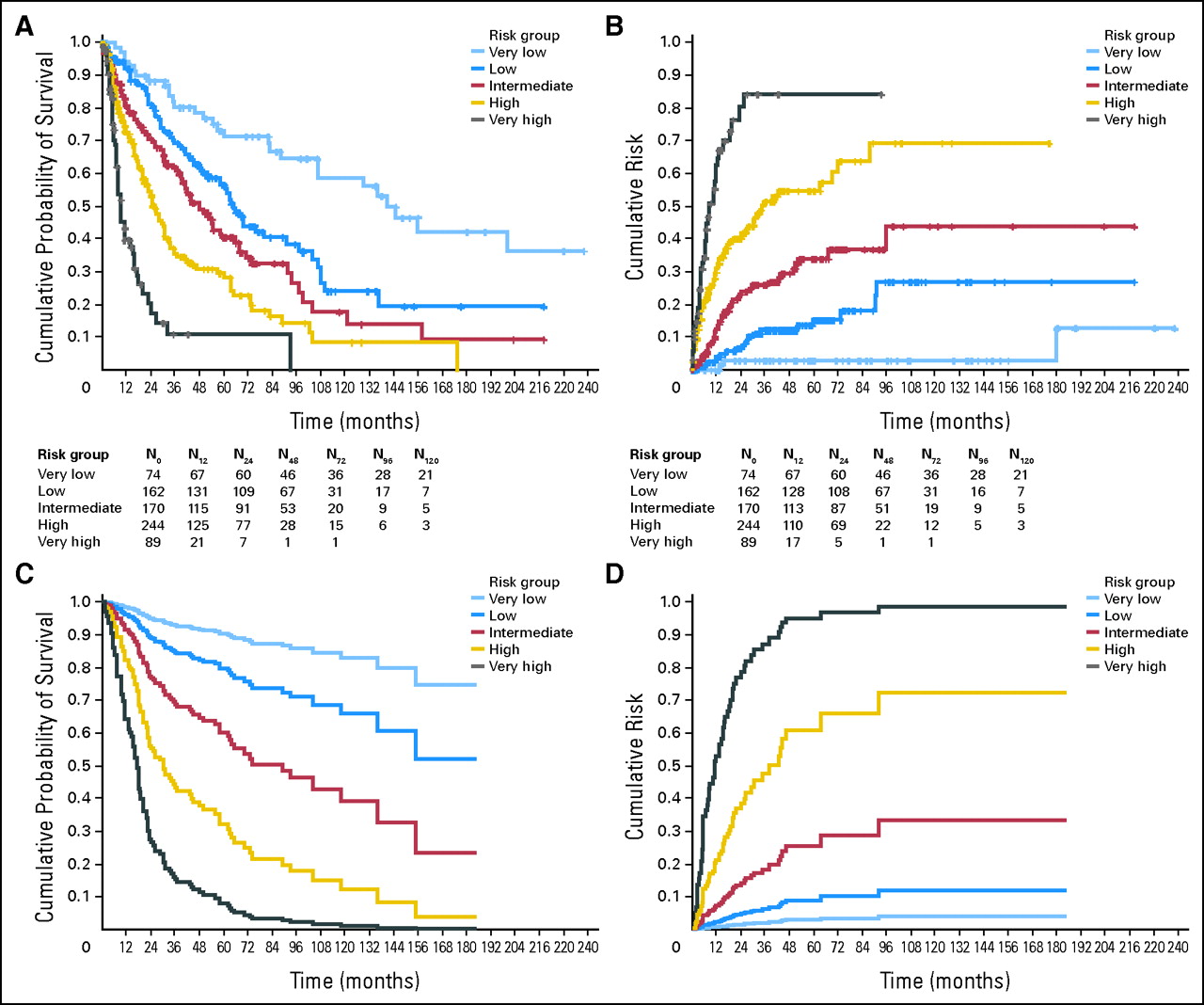
|
|
|
|
|
|
| WHO category | RA, RARS, 5q– | RCMD, RCMD-RS | RAEB-1 | RAEB-2 |
| Karyotype | normal, –Y, del(5q), del(20q) | other abnormalities | complex (>= three abnormalities), chromosome 7 anomalies | - |
| Transfusion requirement | No | at least one RBC transfusion every 8 weeks over a period of 4 months | - | - |
 Copyright © 2001-2014 Daniele
Focosi.
All rights reserved
| Terms of use
| Legal
notices

|
|
|
for |
|
