 TRANSPLANTATION (HSCT / HCT) : intravenous infusion
Table of contents :
|
|
 ))
: easier accessibility and availability, faster engraftment of neutrophil
and platelets when compared to standard bone marrow transplantref.
PBPC collection in children weighing </=25 kg is hampered by technical
and clinical problems related to vascular access, low total blood volume,
anticoagulation, side effects, and psychological impact but large-volume
leukapheresis (LVL) is a safe and effective procedureref
mobilization (30-fold increase) coincides in vivo with a decrease
in BM SDF-1 concentration related to SDF-1 cleavage by marrow serine proteasesref1,
ref2
and dipeptidyl peptidase IV (CD26)ref1,
ref2.
Furthermore, treating HSPCs by pertussis toxin (PT), which causes an interruption
of Gai-protein signaling, did not
affect their BM engraftmentref
but induced their release in the peripheral bloodref,
suggesting that a Gai-protein signaling
identical to that mediated by CXCR4
receptor or other chemokine receptors must be required for HSPC retention
in BM microenvironmentref1,
ref2,
ref3.
Consistent with the hypothesis that the CXCR4/SDF-1 axis plays a major
role in the retention of hematopoietic cells, it has been shown that elevated
numbers of granulocytic precursors were present in the peripheral blood
of mice reconstituted with CXCR4–/– FL cellsref1,
ref2.
The ribosomal
protein SA (RPSA) / 67 kDa laminin receptor (67LR) is a non-integrin
cell-surface receptor with high affinity for laminin which plays a key
role in tumor invasion and metastasis. The role of 67LR in G-CSF-induced
mobilization of CD34+ HSCs from 35 healthy donors was investigated.
G-CSF-mobilized HSCs, including CD34+/CD38-- cells,
showed increased 67LR expression, as compared with unstimulated marrow
HSCs; noteworthy, the level of 67LR expression in G-CSF-mobilized HSCs
significantly correlated with mobilization efficiency. During G-CSF- induced
HSC mobilization, the expression of laminin receptors switched from 6 integrins,
which mediated laminin-dependent adhesion of steady-state human marrow
HSCs, to 67LR, responsible for G-CSF-mobilized HSC adhesion and migration
toward laminin. In vitro G-CSF treatment, alone or combined with
exposure to marrow-derived endothelial cells, induced 67LR up-regulation
in marrow HSCs; moreover, anti-67LR antibodies significantly inhibited
transendothelial migration of G-CSF-stimulated marrow HSCs. Finally, G-
CSF-induced mobilization in mice was associated with 67LR up-regulation
both in circulating and marrow CD34+ cells, and anti-67LR antibodies
significantly reduced HSC mobilization, providing the first in vivo
evidence for 67LR involvement in stem cell egress from bone marrow after
G-CSF administration. In conclusion, 67LR upregulation in G-CSF-mobilized
HSCs correlates with their successful mobilization and reflects its increase
in marrow HSCs, which contributes to the egress from bone marrow by mediating
laminin-dependent cell adhesion and transendothelial migrationref.
A total of 138 patients whose stem cell mobilization failed following chemotherapy
and G-CSF at a dose of 5 mg/kg/d were given
a higher dose of G-CSF (10 mg/kg/d) for
5 days after a 7-day resting period. HSC mobilization was successful in
90 patients, who yielded a median of 3.5x10⁶ CD34+
cells/kg, partially successful in 17 patients (1-2.4x10⁶ CD34+
cells/kg) and failed in the remaining 31 patientsref.
Anyway high-dose G-CSF alone was inefficient as second mobilization regimen
in fludarabin-cyclophosphamide-treated CLL patients who failed to mobilize
after chemotherapy and G-CSFref.
The HSC mobilization-associated granulocytosis severely worsens acute renal
failureref,
in combination with salvage chemotherapy, is a predictable and highly effective
CD34+ stem cells mobilization regimen in pretreated lymphoma
patients. This was an open-label phase II study including 25 pretreated
patients (Hodgkin's disease=4; aggressive NHL=21). The primary end-point
of the study was the successful mobilization of a target cell dose of 2x10⁶
CD34+ cells/kg in lymphoma patients receiving ifosfamide, epirubicin
and etoposide (IEV) chemotherapy and a fixed dose (6 mg) of pegfilgrastim
given as single subcutaneous injection. Following chemotherapy, all patients
had grade 4 neutropenia that lasted a median of 1.5 days (1-3). Pegfilgrastim
treatment was well tolerated and only 2/25 patients required pain-control
medication. CD34+ cells were mobilized in all patients. The
median (range) peak value of peripheral blood CD34+ cells after
IEV chemotherapy and pegfilgrastim was 141x106/L (12.8-386)
and occurred almost invariably on day +14 (13-16). 23 of the 25 patients
underwent a single standard volume leukapheresis to collect a median of
8.7x10⁶ CD34+ cells/kg (1.78-17.3). 24/25 patients
(96%) reached the target cell dose of 2x10⁶ CD34+
cells/kg. High concentrations of circulating CD34+ cells (>
50x106/L) were observed for several days after the achievement
of the peak value. All the study patients were transplanted with their
pegfilgrastim-mobilized CD34+ cells and showed a rapid and sustained
engraftment after high-dose chemotherapyref.
Pegfilgrastim effectively mobilizes PBSC in a poor mobilizer multiple myeloma
patientref
+ SCF
in poor mobilisers : a predictive model [PBSC expected = 0.6+(G-CSF-based
total collection)+2 (rHuSCF-based day 1 collection)] to calculate the cumulative
total of PBSC expected following a maximum of five leukapheresesref1,
ref2.
Failure has been reportedref
+ GM-CSF
: there is no difference between high-dose G-CSF (32 mg/kg/day)
or G-CSF(10 mg/kg/day)+GM-CSF (5 mg/kg/day)ref
+ GHrefref1,
ref2,
ref3
antagonistsref1,
ref2,
ref3,
ref4,
ref5
plasma elevationref1,
ref2
serve as cofactors in cytokine-induced mobilization. Modification of the
endotoxin content by antibiotic treatment may affect the yield of cytokine-induced
mobilizationref
(1.5 g/m2) plus lenograstim
is a safe and effective mobilizing regimenref
(1.8 g/m2 per day for 5 days) + carboplatin
(400 mg/m2 per day for 2 days) + etoposide
(100 mg/m2 per day for 5 days) (ICE) /IL-11
(50-100 mg/kg per day) + G-CSF
(5 mg/kg per day) is successful in mobilizing
large numbers of CD34+ PBSC cells with a limited number (one)
of apheresis collections in patients that have previously been heavily
pretreated with chemotherapy/radiotherapy
(600 mg/m2 twice daily for 10 doses) and etoposide
(200 mg/m2 twice daily for 10 doses (continuous; n=57) or 2
g/m2 over 10 hours on day 5 of etoposide (bolus; n=22)) + filgrastim
(5 mg/kg/d beginning day 14) to mobilize
autologous hematopoietic progenitor cells in 79 patients with high-risk
or relapsed/primary refractory NHL, MM, and Waldenstrom macroglobulinemia.
59% of patients achieved the primary end point (a CD34 cell dose of 5 million/kg
with a single leukapheresis). More bolus etoposide patients achieved the
primary end point (86%) compared with continuous etoposide patients (47%;
P<.0001). The CD34 cell dose collected was greater in bolus etoposide
patients (44 million/kg) than in continuous etoposide patients (10.9 million
per kilogram; P<.0001). Patients took 3 weeks to recover >500/ml
neutrophils and >20000/ml platelets after cyclophosphamide
and etoposide. The overall response rate was 69% for NHL patients and 71%
for MM/WM patients. The TR; was 2.5%. 16% of surviving patients experienced
grade>or=3 nonhematologic toxicity. Patients receiving bolus etoposide
had significantly less grade>or=2 oral mucositis, less use of TPN, and
less need for red blood cell and platelet transfusions. 64 patients (81%)
underwent autologous HSCT, with prompt engraftment. 4 patients (5%) did
not undergo autologous HSCT because of toxicity from high-dose cyclophosphamide
and etoposideref
Both T cells and natural killer (NK) cells from HCT donors have been
implicated in the graft-versus-tumor (GVT) effects after allogeneic HCT
(Truitt & Atasoylu, 1991; Glass et al, 1996; Giebel et al, 2003). The
GVT effect of T cells has been indirectly confirmed by the results of T-cell
depletion trials (Link, 1999; Barge et al, 2001; Chakraverty et al, 2001).
Preclinical studies in the canine HCT model showed that grafts created
their own marrow space via subclinical graft-versus-host (GVH) reactions,
thereby ensuring stable engraftment (Storb et al, 1999). Data from clinical
trials in patients showed that a low degree of donor T-cell chimaerism
on day +28 after non-myeloablative HCT was associated with an increased
risk of rejection and that high donor T-cell chimaerism was beneficial
in mediating GVT effects (Antin et al, 2001; McSweeney et al, 2001; Maris
et al, 2003; Niederwieser et al, 2003). The level of donor T-cell engraftment
(chimaerism) is currently being used as a surrogate marker for donor engraftment
after non-myeloablative HCT (Childs et al, 1999; Antin et al, 2001; McSweeney
et al, 2001).
This study confirms the substantial variation in PBSC cellular subset
composition as reported in previous studies of conventional myeloablative
allogeneic HCT recipients (Dreger et al, 1994; Ottinger et al, 1996). Our
analysis showed positive correlations between almost all cellular subtypes
within the PBSC product.
Results from this study are the first published indication in the truly non-myeloablative setting that infused quantities of some cellular subsets of PBSC products from HLA-matched siblings are associated with important post-HCT outcomes of engraftment (T-cell chimaerism) and survival. The cellular components that were associated with higher donor T-cell chimaerism on day +28 were TNC and CD56+ cell numbers. Given the observation that TNC numbers were highly correlated with CD3+ cell numbers in this study, the relationship of TNC on donor T-cell chimaerism possibly reflected the known role of T cells in establishing donor engraftment after myeloablative and non-myeloablative HCT (Champlin, 1993; Drobyski, 2000; Kreiter et al, 2001). NK and NKT cells analysed separately were marginally associated with T-cell chimaerism; however, the association was stronger when the two values were combined. CD56 has been used as a surrogate marker to describe NK cells, and it was known that NK-cell alloreactivity could influence engraftment and relapse rates after HLA-mismatched myeloablative HCT (Bornhauser et al, 1999; Ruggeri et al, 1999, 2001). This alloreactivity was thought to be the result of the missing expression of HLA-class I epitopes on haematopoietic cells (Ciccone et al, 1992). Whether donor NK cells can exert anti-host activity after HLA-identical related donor HCT is presently unknown (Dupont & Hsu, 2004). Interestingly, a recent report showed that patients who rejected their unrelated grafts after a non-myeloablative conditioning regimen had significantly lower donor NK-cell chimaerism compared with patients who did not experience rejection (Bornhauser et al, 2001). However, it was unclear if, and to what level, these patients were HLA-matched in this report, and no data were given about the actual number of donor cells transplanted.
We suspected that certain cellular subsets in PBSC grafts would be associated with better progression-free and overall survival after non-myeloablative HCT, as this approach relies mainly on graft anti-tumour responses mediated by donor cells (Barrett & Malkovska, 1996; Appelbaum, 2001). In fact, only B cells and CD34+ cells were associated with improved outcomes. Specifically, higher doses of B cells appeared to be associated with a greater occurrence of chronic GVHD, better progression-free survival, and improved overall survival, while the effect of higher numbers of CD34+ progenitor cells was associated only with better overall survival. The finding that higher B-cell numbers were associated with an increase in chronic GVHD in the current study was unexpected, two previous reports had shown that high numbers of CD34+ cells were associated with an increased risk of chronic GVHD after both myeloablative and reduced-intensity-conditioning HCT (Zaucha et al, 2001; Perez-Simon et al, 2003). The association of B cells with chronic GVHD is also the probable explanation for the positive effect of B cells on progression-free and overall survival found in this study, possibly through GVT effects. In addition, humoral responses of B cells against malignant host cells, which have been described for potential CML-associated antigens (Ling et al, 1998; Wu et al, 2000), might have been responsible for the anti-tumour responses. However, it seems most likely that the donor CD20+ cells could have functioned as antigen-presenting cells (Chesnut & Grey, 1986; Pernis & Weber, 1989), thereby facilitating the donor T cells to exert more GVT and GVHD effects.
Higher numbers of CD14+ cells (monocytes) were associated with worse overall survival in our analysis. As CD14+ cells in PBSC might decrease allogeneic T-cell responses (Mielcarek et al, 1997), one might speculate that GVT effects may have been diminished in patients who received a higher CD14+ cell dose in the PBSC graft.
As expected, patients categorized before HCT as having aggressive malignant disease had a higher risk for disease relapse/progression and worse progression-free and overall survival. These results confirmed the prognostic value of this clinical categorization, similar to the high dose conventional HCT setting; patients classified as having aggressive diseases had worse outcomes after non-myeloablative conditioning as well.
Another finding consistent with previous studies was that patients who
had received 2 Gy TBI only as conditioning showed lower donor T-cell chimaerism
on day 28 compared with patients who had received additional fludarabine
or a combination of autologous and allogeneic transplantation (McSweeney
et al, 2001). As fludarabine is known to be immunosuppressive and to enhance
the effect of radiation (Gregoire et al, 1999; Van Den Neste et al, 2001),
it is likely that additional suppression of host immune-competent cells
caused by fludarabine enabled the achievement of more rapid donor T-cell
chimaerism. Our data were consistent with previous studies showing that
donor T-cell chimaerism can be used as a surrogate marker for engraftment
(Childs et al, 1999; Antin et al, 2001; McSweeney et al, 2001; Niederwieser
et al, 2003), although there were too few rejections in this set to confirm
this hypothesis.
In summary, we have shown that the doses of CD34+ cells, B cells and
monocytes in the HLA-matched related PBSC graft influenced outcomes after
non-myeloablative HCT and confirmed that variations in conditioning regimens,
disease status, and age of donors and recipients were important. Evaluations
in patients receiving PBSC from alternative donors (e.g. HLA-matched unrelated
or HLA-mismatched donor recipient pairs) are needed to support and further
exploit these findings. Furthermore, prospective clinical studies with
larger numbers of patients will be required. If these results are confirmed,
then clinical studies evaluating graft engineering for selective cellular
enrichment or depletion may be important in improving clinical outcomes
after non-myeloablative conditioning for allogeneic HCT.
This process mimics enhancement of the physiological release of stem
cells and progenitors from the bone marrow (BM) reservoir in response to
stress signals during injury and inflammation. Currently, mobilized cells
are the preferable and major source of stem and progenitor cells harvested
for autologous and allogeneic transplantations because of the higher yield
of these cells, leading to faster engraftment and decreased procedural
risks compared with harvested BM cells. The emerging picture of stem cell
mobilization involves interfering with the physiological interplay between
mesenchymal stromal and hematopoietic cells regulating both bone and BM
remodeling processes, which also mediate stem/progenitor cell proliferation
and migration. The mobilization process is initiated by stress-induced
activation of neutrophils and osteoclasts by chemotherapy and repeated
stimulation with cytokines such as G-CSF, resulting in shedding and release
of membrane-bound SCF, proliferation of progenitor cells, as well as activation
and/or degradation of adhesion molecules such as VLA-4 and P/E selectins.
The dynamic secretion and inactivation of the chemokines SDF-1/CXCL12 and
IL-8/CXCL8, and multiple cycles of inactivation and degradation of BM components
by proteolytic enzymes, such as elastase, cathepsin G, proteinase 3, CD26,
and various MMPs, are implicated as major players in progenitor mobilization.
These mechanisms will be reviewed in this section. The road to stem cell
mobilization began in the 1960s, with publication of a few reports documenting
the presence of hematopoietic stem cells in the peripheral blood of mice,
dogs, and monkeys, followed by reports revealing low levels of progenitors
in the human circulation during steady-state homeostasis. However, steady-state
progenitors in the circulation of mice were shown to be inferior to BM
as a source of repopulating stem cells. More important, although sporadic
clinical transplantations documented successful hematopoietic recovery,
in most cases graft failure was detected in autologous, syngeneic, and
fully matched related allogeneic transplantations, using steady-state peripheral
blood cells. Moreover, the first successful human blood leukocyte transplantations
were not better than BM as a source of repopulating stem cells (reviewed
in Korbling and Fliender (Korbling M, Fliender TM. History of blood stem
cell transplants. Blood stem cell transplants. In: Gale RP, Juttner CA,
Henon P eds. Peripheral blood stem cell autographts. New York: Cambridge
University Press; 1994:9) and To et alref).
While the levels of human progenitors in the circulation during steady-state
homeostasis are low, they are significantly elevated in patients with myeloproliferative
disordersref.
Similarly, a transient increase in circulating progenitors was documented
in dogs treated with dextran sulfate and humans treated with endotoxin
or other stress-inducing mobilization agents. These encouraging results
were followed by preliminary reports in the late 1970s documenting increased
levels of progenitors in the circulation of patients after chemotherapy
treatment with cyclophosphamide and other drugs. Technical improvements
including in vitro colony assays and the availability of large-scale
harvesting of human progenitors by continuous-flow leukopheresis enabled
closer examination of chemotherapy-/druginduced mobilization in treated
patientsref
(Korbling M, Fliender TM. History of blood stem cell transplants. Blood
stem cell transplants. In: Gale RP, Juttner CA, Henon P eds. Peripheral
blood stem cell autographts. New York: Cambridge University Press; 1994:9).
In the early 1980s, To and colleagues reported high levels of progenitors
in the circulation of leukemia patients in early remissionref.
Clinical studies by this group were the first to document the beneficial
faster repopulation in patients transplanted with autologous mobilized
peripheral blood (PBL). Initially, the mobilization protocols were based
on chemotherapy alone. However, after the discovery of human G-CSF by Welte
et alref,
mobilization protocols began to include G-CSF, which today is the standard
mobilizing agentref.
During late embryonic development, both human and murine stem cells migrate
via the blood circulation from the fetal liver to the BM and repopulate
this tissue with high levels of immature and maturing cells. These in turn
are released into the circulation, while a small pool of undifferentiated
stem and progenitor cells within the BM is maintained throughout life.
Prior to their localization in the murine fetal liver, hematopoietic stem
cells can be isolated from the aorta-gonad-mesonephros (AGM) region and
the yolk sac even before completion of blood circulation developmentref.
However, some of these stem cells are not definitive, since they cannot
rescue and repopulate lethally irradiated recipients because of their inability
to migrate in the host circulation and home to and repopulate the BM. These
predefinitive stem cells isolated from the murine yolk sac can still engraft
the liver of newborn mice preconditioned as fetuses with chemotherapy if
the predefinitive stem cells are directly injected into this organref.
Furthermore, an additional maturation step provided to predefinitive progenitors
isolated from the AGM region, in the form of in vitro incubation on stromal
cells and stimulation with cytokines, shifts these cells into definitive
stem cells with functional migration, homing, and repopulation potentialref.
Murine embryos that lack the chemokine SDF-1 or its receptor CXCR4 have
multiple lethal defects, including impaired seeding of the fetal BMref.
The essential roles of SDF-1/CXCR4 interactions has been shown in a functional,
preclinical model for human stem cells using non-obese diabetes severe
combined immune deficient (NOD/SCID) mice as recipients. Homing and repopulation
in both primary and serially transplanted mice with human CD34 stem cells
was dependent on CXCR4 signalingref1,
ref2,
which is dynamically expressed by human progenitorsref.
Moreover, SDF-1-mediated migration of human CD34 cells in vitro correlated
with their in vivo repopulation potential in transplanted patientsref.
In conclusion, stem cell motility (migration, homing, and release) is essential
for BM repopulation and for the development of the hematopoietic system.
The role of circulating stem cells in steady-state homeostasis is currently
unknown. While some progenitors are needed for seeding of the thymus, which
requires migration of lymphocyte precursors from the BM, this process is
markedly reduced in adult liferef.
Stem cells migrate also to nonhematopoietic organs such as the liver, especially
during liver injury/inflammation, creating an alarm situation and transmitting
stress signals that mobilize and recruit stem cells as part of organ repairref.
Ramshaw et al suggest that circulating stem cells can reengraft the BM,
documenting successful engraftment of unconditioned murine recipients transplanted
with high doses of BM cellsref.
In support of this approach, Wright et al elegantly demonstrated rapid
clearance of intravenous transplanted mouse stem cells from the murine
circulation within a few minutes. By using genetically marked parabiotic
mice with a shared circulation, they showed that the low levels of circulating
stem cells can durably reengraft the BM, suggesting continuous release
and reengraftment of the BM as sequential events taking part in physiological
processesref.
In parallel, Abkowitz et al, using the same parabiotic model, revealed
only low levels of reengrafted stem cells with secondary recipient repopulation
potential (1–2.5%) in the partner BM as opposed to high levels of progenitors
in the spleen. These investigators suggest the release of stem cells into
the circulation functions also as an apoptotic pathway for the BM to regulate
excess amounts of progenitors because of limited numbers of stem cell nichesref.
Of interest, these investigators also applied G-CSF- and SCF-induced mobilization
to the parabiotic mice pairs, documenting a dramatic 4- to 10-fold increase
(from 1–2.5% to 10.1%) in the levels of reengrafted stem cells with secondary
repopulation potential in the partner BM. These results demonstrate that
inducing the release of stem cells in response to stress signals also increases
their homing back and reengraftment of the stimulated/stressed BM, confirming
the repopulation potential of mobilized progenitors harvested for clinical
protocols. These results also support the hypothesis that tissue repair
of damaged BM can be supported by intensified homing and reengraftment
of circulating hematopoietic stem cells, in response to stress signals,
which is similar to migration of mature leukocytes to sites of inflammation
as part of the immune system host defense. The concept of BM niches occupied
by stationary, quiescent stem cells may be an oversimplification of a more
dynamic situation. Higher proliferative and migratory turnover of definitive,
repopulating stem cells involves their release into the circulation, migration,
homing, and reengraftment of the BM as part of their physiological development.
Bradford et al revealed that even the most primitive murine stem cells
in the BM must cycle at least once every 30 daysref.
Proliferation and release of hematopoietic stem and progenitor cells require
dynamic cycles of BM destruction/restructuring, which seem to be linked
to bone remodeling by osteoclast/osteoblast interactions. Moreover, both
processes are likely to be orchestrated by the same factors. For example,
continuous G-CSF treatment to prevent neutropenia has also been shown to
induce osteoporosis in some juvenile patients with severe congenital neutropeniaref
and in transgenic mice that overexpress this cytokineref.
More important, there is a dramatic increase in the number of murine osteoclasts
in response to repetitive G-CSF stimulation, leading to osteoclast-mediated
bone resorption during stem cell mobilization and calcium release, which
can induce detachment of hematopoietic progenitors from fibronectin
in vitroref.
Unexpectedly, treatment of mice with pamidronate, an inhibitor of osteoclast-mediated
bone resorption, together with G-CSF increased the levels of mobilized
progenitors in the murine circulationref.
Osteoclasts secrete high levels of IL-8, a mobilization-inducing chemokineref1,
ref2.
Another chemokine, SDF-1, which also participates in G-CSF–mediated mobilization,
induces osteoclasts to secrete the metalloproteinase MMP-9ref.
This enzyme is involved in stem cell mobilization by shedding the membrane-bound
cytokine SCF within the BM and releasing this cytokine into the circulationref.
MMP-9 also induces migration of osteoclasts to the cortical bone, adjacent
to the SDF-1 and stem cell–rich endosteum region, to form resorption pitsref1,
ref2.
These results suggest that the role of osteoclasts in mobilization could
be related to the rapid increase in their numbers and to the secretion
of chemokines and proteolytic enzymes. Activation and secretion of proteolytic
enzymes lead to cleavage and degradation of the BM extracellular matrix,
adhesion molecules, cytokines, and chemokines and therefore facilitate
transendothelial migration and stem cell release from the BM.7 These processes
mediate both peripheral bone and medullar BM remodeling as well as stem
cell proliferation and release by inactivating the BM microenvironment.
Subsequently, stem cells migrate via the circulation, home back to the
BM, and repopulate the damaged/destructured sites in this organ as part
of tissue repair and the continuous replenishment of the blood with new
immature and maturing hematopoietic cells of all lineages, while maintaining
a small pool of undifferentiated stem and progenitor cells within their
renewed BM niches. In support of this hypothesis, by applying complementary
DNA array technology, 2 recent studies revealed high expression of the
proteolytic enzyme proteinase 3 and low expression of a proteinase 3 inhibitor
in isolated human BM CD34 cells compared with enriched mobilized CD34 cells
isolated from G-CSF-treated healthy donorsref1,
ref2.
These results suggest that proteinase 3, with its ability to cleave connective
tissue in the BM, is needed for maintaining a niche for hematopoietic stem
and progenitor cells. Recent findings document that proteinase 3 also cleaves
the cell cycle checkpoint p21, inducing cells to enter the cell cycleref.
This could explain the higher levels of cycling BM CD34 cells compared
with the more quiescent mobilized PBL CD34 cellsref1,
ref2.
Immature mesenchymal bone-forming osteoblasts lining the stem cell–rich
endosteum region secrete high levels of SDF-1ref.
In response to stimulation with this ligand, hematopoietic stem cell–derived
osteoclasts secrete MMP-9ref1,
ref2.
Thus, peripheral periosteal bone and medullar BM destruction and remodeling
are sequential events mediated by the same factors, i.e. MMP-9, G-CSF,
SCF, IL-8, and SDF-1, which regulate both new bone formation and stem cell
self-renewal, migration, and development. Applying single or multiple stress
signals interferes with steady-state homeostasis, creating an alarm situation
leading to increased release of immature and maturing hematopoietic cells
from the BM into the blood circulation, which occurs naturally during inflammation
and injury. For example, increased levels of G-CSF and other cytokines
secreted by long-distance runners during marathons cause blood leukocytosis,
in particular higher levels of CD34/CD38+ committed progenitor
cells, but not more primitive CD38- stem cells compared with
control individuals, reflecting adaptation of BM activity to stress induced
by extensive exerciseref.
Increased cell release from the BM reservoir is part of the immune system
host defense during inflammation as a result of infection- or injury-mediated
release of stress signals. This release is induced clinically or in animal
models by a wide range of molecules and/or treatments: DNA damaging, single
chemotherapeutic drugs such as cyclophosphamide,
or combined chemotherapy regimens such as iphosphamide,
carboplatin and etoposide (ICE)
and etoposide, methylprednisolone,
ara-c and cisplatin (ESHAP);
cytokines such as G-CSF, GM-CSF, SCF, and flt-3 ligand; and chemokines
such as IL-8, Mip-1, Groß, and SDF-1ref.
These molecules differ in their mode of administration, the time frame
needed to achieve mobilization, the type of cells mobilized, and the efficiency.
G-CSF, the most commonly used mobilization agent, is usually administered
daily at a dose of 5–10 µg/kg for 5–10 days, alone or after chemotherapyref.
However, a substantial number of patients, especially patients having extensive
chemotherapy that leads to BM aplasia, older patients, and a minority of
healthy individuals, are poor mobilizersref.
For example, increasing age and longer intensive chemotherapy treatment
in old multiple myeloma patients inversely correlate with successful mobilizationref.
These patients are usually treated with higher doses of G-CSF, GM-CSF followed
by G-CSF, or a combination of G-CSF and SCF. Recently, it was reported
that some chronic
myeloid leukemia (CML)
patients treated with high doses of imatinib (Glivec, STI 571) suffer hematological
toxicity and can develop drug-induced neutropeniaref.
In addition, some treated patients failed to mobilize well, requiring discontinuation
of the drug during the mobilization process and the addition of SCF to
G-CSF, to successfully overcome the poor mobilizationref.
However, while SCF stimulation increases the levels of mobilized CD34+
progenitors, this treatment also has side effects such as allergic reaction
due to activation of mast cells. These studies stress the need to identify
and characterize the mechanisms of stem cell mobilization in order to develop
better strategies for poor mobilizers. Chemotherapy followed by repetitive
G-CSF–induced mobilization is a multistep process. Anchorage of stem and
progenitor cells to the BM microenvironment by activation of adhesion interactions,
which are facilitated by membrane-bound SCF or SDF-1, needs to be disruptedref.
Papayannopoulou elegantly demonstrated that the integrin VLA-4 plays a
critical role in stem cell mobilization since anti-VLA-4 antibodies prevent
adhesion molecules expressed on the surface of hematopoietic progenitors
from binding their ligand VCAM-1 expressed by stromal cells. This induces
rapid mobilization of primate and murine stem cells, which also involves
signaling via the c-kit receptor. Moreover, as a result of inactivation
during the mobilization process, mobilized CD34+ cells have
lower levels of VLA-4 and c-kit expression compared with BM progenitorsref1,
ref2.
In addition, sulfated polysaccharides such as Fucoidan lead to rapid mobilization
of progenitorsref
since they compete for adhesion interactions with selectins and for SDF-1
binding to the BM endothelium via its nonsignaling C-terminus, inducing
inactivation of selectin interactions as well as release of SDF-1 into
the circulation. Levesque et al revealed increased release of elastase
and cathepsin G within murine BM during Cy- and G-CSF–induced mobilization;
peak levels were reached during stem cell egressref.
These enzymes cleaved VCAM-1 expressed by stromal cells, preventing adhesion
of progenitors via VLA-4 as part of the mobilization process. In addition
to their distinctive adhesive properties, mobilized CD34 progenitors differ
from their BM counterparts in several ways. Besides reduced VLA-4, c-kit,
and CXCR4 expression, a significantly higher percentage of mobilized progenitors
are noncycling quiescent cellsref1,
ref2.
Prior to mobilization, hematopoietic progenitors proliferate within the
BM, and this feature could involve proteinase 3–mediated cleavage of p21
in response to neutrophil stimulationref.
Mobilized progenitors have higher levels of the proapoptotic genes caspase
3, 4, and 8 and reduction in inhibitors of apoptosis such as antiproteinase
2, compared with human BM CD34 cellsref,
supporting the hypothesis of Abkowitz et al that release into the circulation
may also serve as an apoptotic pathway for stem cellsref.
Enhanced repopulation documented with mobilized progenitors is due to significantly
higher cell doses, in particular committed progenitors, which lead to faster
neutrophil and platelet recoveryref.
However, better leukemia-free survival and overall survival were recently
documented in acute
myeloid leukemia (AML)
patients transplanted with increased cell doses of matched BM cells compared
with lower doses of BM cells and more important, also compared with high
cell doses of matched mobilized PBL, suggesting BM as a superior source
of stem cells for HLA-identical allogeneic transplantationsref.
New protocols aimed at increasing the levels of long-term repopulating
stem cells as the major source while maintaining short-term repopulating
cells need to be developed. Mobilized human CD34 progenitors express reduced
levels of the SDF-1 receptor CXCR4, which correlates with improved mobilizationref1,
ref2,
suggesting involvement of SDF-1/CXCR4 interactions in the mobilization
process. Overexpression of SDF-1 in the murine circulation leads to stem
cell mobilizationref.
In functional preclinical animal models for human stem cells such as preimmune
sheep and immune-deficient NOD/SCID mice, mobilized CD34+ cells
are inferior in their repopulating potential compared with similar cell
doses of CD34 cells obtained from human BM. This is most probably due to
their reduced levels of surface CXCR4, which is needed for SDF-1–mediated
directional homing and repopulation in transplanted mice and increased
levels of short-term repopulating cellsref.
These results should be interpreted with caution, since short-term stimulation
with human SCF upregulated surface CXCR4 expression on human CD34+
cells and murine SCF is much less potent in stimulating human progenitorsref1,
ref2.
In addition, other factors such as complement C3a improve SDF-1–mediated
directional migration of human progenitors and the in vivo homing of murine
progenitors. However, some of these factors can be species-specific or
absent in immune-deficient miceref.
Of interest, priming of mobilized human progenitors by several inflammatory
molecules improves their chemotactic responses to SDF-1 (Wysoczynski M,
Reca R, Kucia M, et al. Mobilized peripheral blood stem/progenitor cells
are primed by several inflammatory molecules for their chemotactic responses
to sdf-1: a molecular explanation as to why mobilized peripheral blood
cells engraft faster than bone marrow cells after transplantation. Paris,
France -July 5–8, 2003; 32nd Annual Scientific Meeting of the International
Society for Experimental Hematology (ISEH). abst 277. 2003). We have demonstrated
increased levels of SDF-1 production in the murine BM in response to DNA-damaging
chemotherapy drugs, including Cy and 5-fluorouracil (5-FU), most probably
to prevent cell death since this chemokine is also a survival factor for
stem cellsref1,
ref2,
ref3.
Rafii and Hattori et al demonstrated that the increased levels of SDF-1
in response to stress-mediated 5-FU mobilization in mice induce release
of MMP-9, which cleaves and releases membrane-bound SCF, leading quiescent
stem cells to proliferate in the BM prior to their mobilization into the
circulationref.
SDF-1 mediates secretion of MMP-2 and MMP-9 from human CD34+
cellsref,
and these proteolytic enzymes inactivate SDF-1 by cleaving a few amino
acids in the N terminusref.
We demonstrated that each injection of G-CSF stimulates mesenchymal cells
such as immature osteoblasts to secrete SDF-1, leading to a transient increase
in SDF-1 levels within the BM. This oscillating increase is followed by
a profound decrease due to inactivation by proteolytic enzymes, mostly
by neutrophil elastase, with the lowest levels of this ligand released
during cell mobilizationref.
Levesque et al reported that in addition to inactivation of BM SDF-1 by
elastase and cathepsin G, these proteolytic enzymes can also cleave part
of the CXCR4 receptor N terminus in the BMref,
partially inactivating SDF-1 signaling and directional migration. Of interest,
neutrophils secrete the proteolytic enzymes elastase, cathepsin G, and
proteinase 3 in response to G-CSF, and neutrophil elastase also inactivates
G-CSFref,
which is essential for regulating release of neutrophils from the BM. However,
expression of G-CSF receptors by neutrophils is not required because of
an indirect mechanism that involves proteolytic reduction of SDF-1 within
the murine BMref.
Of interest, the central role of elastase in regulating release of BM leukocytes
is suggested by inherited genetic mutations in the elastase gene, leading
to 2 forms of neutropenia in juvenile patients (severe congenital neutropenia,
or Kostmann disease, and cyclic neutropenia) that require continuous treatment
with G-CSFref.
CD26, another proteolytic enzyme that inactivates SDF-1, is expressed by
human CD34 progenitors and is involved in G-CSF–induced mobilization in
miceref.
A role for CXCR4 signaling in cell egress from the BM to the circulation
emerges from several studies. In parallel to gradual reduction of BM SDF-1
during G-SCF administration, CXCR4 expression is elevated within the human
and murine BM, reaching peak levels at the time of mobilizationref.
While some CXCR4 inhibitors also lead to release of human progenitorsref,
and treatment with pertussis toxin, which blocks Gi-mediated signaling
in 7 transmembrane G coupled receptors such as CXCR4, induced mobilization
in miceref,
SDF-1/CXCR4 interactions are also needed for cell egress. Treatment of
mice with G-CSF and neutralizing anti-CXCR4, anti-SDF-1 Ab, or inhibitors
of CD26 reduced the mobilization levelsref1,
ref2.
Finally, warts, hypogammaglobulinemia, immunodeficiency and myelokathexis
(WHIM) syndrome, a rare inherited immunodeficiency disease associated with
mutations in CXCR4, is characterized by neutropenia and B-cell lymphopenia.
In one case, an affected individual was born with cardiac malformation,
resembling the murine model in which knocking out CXCR4 or SDF-1 results
in a lethal phenotype associated with lack of BM seeding, B-cell development,
and cardiac septum formationref.
Thus, SDF-1/CXCR4 interactions may have a role in the regulation of the
routine and active egress of progenitor and maturing cells from the BM
into the circulation. Taken together, these results decipher key mechanistic
insights into stress-induced mobilization that mimic and amplify naturally
occurring recruitment of progenitors during alarm situations. We suggest
the BM as a reservoir for immature and maturing hematopoietic cells to
be released into the circulation upon stress signals, to migrate to injured
sites, and to contribute to host defense and tissue repair. The steady-state
balance in the BM is disrupted, leading to transient increased production
of SDF-1, and proliferation and activation of neutrophils and osteoclasts.
Release of proteolytic enzymes is followed by shedding of membrane-bound
SCF, proliferation of hematopoietic progenitors, increasing surface CXCR4
expression and inactivation of SDF-1, G-CSF, the BM adhesion machinery,
and extra cellular matrix (ECM). These events are intensified in each cycle
of stimulation by G-CSF, eventually leading to release of progenitors into
the circulation. A better understanding of the process by which progenitors
egress from the BM will eventually lead to the development of improved
mobilization protocols, in particular for patients who are poor mobilizers.
are believed to be pluripotent and generally express CD34 on their surfaceref.
A commercial assay has now been developed for this intracellular enzyme
(Aldecount, Stemco Biomedical, Durham, NC). As this assay detects both
CD34+ and CD34- cells, and appears to differentiate
viable from non-viable cells (Balber A, Gentry C, Pritchard C, Baucom C,
Deibert E, Smith C. ABC-mediated efflux of ALDH reaction product: implications
for enumeratioin and isolation of blood progenitor cells. Biol Blood Marrow
Transplant. 2003;9:231a), its use may eventually lead to a major change
in how HPCs are enumerated in transplant grafts.
has become the standard against which all other mobilization agents are
measured. This is because it has been shown to both mobilize more CD34+
cells and have less toxicity than any other single agent against which
it has been tested to date. It is not completely without toxicity, however,
given that there have been deaths attributed to thrombosis (acute myocardial
infarction and stroke) in sibling donorsref,
possibly related to receptors on platelets for G-CSFref.
Also, a recent study of serial ultrasounds in donors receiving G-CSF prompted
by 4 reports of splenic rupture related to G-CSF demonstrated universal
enlargement of the spleen during mobilization, with regression of size
after discontinuing the growth factorref1,
ref2.
Other effects of G-CSF which are shared with GM-CSF include pain, nausea,
vomiting, diarrhea, insomnia, chills, fevers, and nightsweatsref1,
ref2
as a single agent is used less often today for mobilization than G-CSF,
because it mobilizes somewhat less well than G-CSFref
and because of a relatively higher incidence of both mild and severe side
effectsref.
However, the fact that it can be more cost effective, and that there are
reports of improved immune reconstitution with GM-CSF relative to G-CSFref1,
ref2,
ref3,
ref4,
has caused some clinicians to reconsider its use. Further, for the patient,
or normal donor who has failed to mobilize adequately on G-CSF alone, the
combination of GM-CSF with or followed by G-CSF has been shown to be efficaciousref1,
ref2,
ref3.
(Law P, Young D, Peterson S, Lane T, Ho A. Mobilization and collection
of peripheral blood progenitor cells from normal subjects treated sequentially
with GM-CSF and G-CSF. Blood. 1996;88:1578a),
now commonly used among cancer patients undergoing chemotherapy to maintain
hemoglobin in the near normal range, also has some ability to mobilize
CD34+ cellsref.
In the hard to mobilize patient, its use may therefore be doubly beneficial.
and darbopoietin
are now available and are in clinical trials as mobilizing agents. They
have the benefit of very long half-lives and so add an important measure
of patient convenience and the probability that timing of collection may
be more flexible without sacrificing optimal collections.,
is currently in clinical trials as a mobilizing agent. It is the first
agent to be tried for mobilization based on a rational understanding of
its mechanism of action relative to HPC-stromal cell interactions. While
it mobilizes CD34+ cells adequately on its own, it significantly
improves the mobilization capacity of G-CSF when used in combination with
G-CSF in mice. Clinical trials in humans with various diseases are in progress,
including myeloma.
and sargramostim
have been approved by the Food and Durg Administration (FDA) for use as
autologous stem cell mobilizing agents. Thus, these have been the only
cytokines used to mobilize allogeneic PBSCs. The majority of the initial
Phase 2 studies using mobilized PBSCs in an allogeneic setting utilized
G-CSF (10–16 µg/kg/day for 5 days). Leukapheresis was performed on
day 4 or day 5 after G-CSF treatment. Fischmeister et alref
followed CD34+ in the peripheral blood after either G-CSF or
GM-CSF treatment and showed that CD34+ cells peaked in the blood
between days +4 and +5 for G-CSF and days +5 and +6 after GM-CSF treatment.
Data from Seattle suggested that larger doses of G-CSF (16 µg/kg/day)
may result in even higher CD34 yields at the time of pheresis on day 5ref.
We have assessed the effect of 5 days of of G-CSF (10 µg/kg) on the
numbers of leukocyte subsets in the peripheral blood of 100 consecutive
normal allogeneic PBSC donors. It is clear from these data that G-CSF has
a pleiotropic effect of increasing the numbers of circulating neutrophils
and monocytes (WBC) as well as T cells (both CD4 and CD8), NK cells and
B cells. Korbling and Anderlini compared the allograft content after G-CSF
mobilization to cellular contents of BM harvests. These data suggest a
3- to 4-fold enhancement of CD34+ cells and a 10- to 20-fold
increase in the number of CD3+ T cells in PBSC products compared to BM
harvestsref|
|
n |
|
|
|
|
|
|
||||||
|
|
|
|
|
|
|
|
|
|
|
|
|
||
| Vigoritoref | 37 | 16 | 18 | 12 | 17 | 78 | 63 | 27 | 19 | 100 | 50 | 47 | 51 |
| Blaiseref | 101 | 15 | 21 | 13 | 21 | 23 | 21 | 44 | 42 | 50 | 28 | 61 | 61 |
| Powlesref | 39 | 17.5 | 23 | 11 | 18 | 31 | 35 | 68 | 58 | 44 | 40 | 70 | 68 |
| Heldalref | 61 | 17 | 23 | 13 | 21 | 17 | 10 | 21 | 10 | 56 | 27 | 80 | 73 |
| Schmitzref | 350 | 12 | 15 | 15 | 20 | ND | ND | 52 | 39 | 74 | 53 | ND | ND |
| Coubanref | 228 | 19 | 22 | 16 | 22 | 7.5 | 16 | 40 | 40 | 71 | 55 | 68 | 55 |
| Bensingerref | 172 | 16 | 21 | 13 | 19 | 21 | 30 | 64 | 57 | 46 | 35 | 66 | 54 |
|
|
|
|
|
|
| # donors | 96 | 102 | 32 | |
| Total # LP procedures required | 1.26 ± 0.5 | 1.20 ± 0.5 | 1.6 ± 0.7 | G vs G/GM: .5; G vs GM: .002 |
| CD34 (x 106/kg) | 9.8 ± 7.2 | 10.9 ± 7.9 | 3.7 ± 2.0 | G vs G/GM: .28; G vs GM: < .001 |
| CD3 (x 108/kg) | 3.5 ± 2.4 | 1.8 ± 1.5 | 1.8 ± 1.8 | G vs G/GM: < .0001; G vs GM: < .001 |
| CD4 (x 108/kg) | 2.3 ± 1.4 | 1.3 ± 1.0 | 0.9 ± 0.8 | G vs G/GM: < .0001; G vs GM: < .001 |
| CD8 (x 108/kg) | 1.1 ± 0.7 | 0.4 ± 0.4 | 0.3 ± 0.26 | G vs G/GM: .56; G vs GM: .002 |
| CD19 (x 108/kg) | 0.97 ± 0.9 | 0.89 ± 0.63 | 0.45 ± 0.4 | G vs G/GM: .56; G vs GM: .002 |
| CD16/56 (x 107/kg) | 4.63 ± 3.7 | 2.98 ± 2.36 | 1.66 ± 1.18 | G vs G/GM: < .001; G vs GM: < .0001 |
|
|
|
|
|
|
| acute grade 2–4 | 51 | 65 | 13 | G vs GM: .003
GM vs G/GM: < .0001 G vs G/GM: .11 |
| acute grade 3–4 | 11 | 13 | 0 | .07 |
| chronic GVHD | 86 | 88 | 65 | NS |
| survival (2 years) | 62
high 38 low 88 |
56
high 28 low 5 |
59
high 35 low 69 |
NS |
at its position Pro2. CXCL12/SDF-1a
induces migration of HSCs and progenitor cells, and it is thought that
CXCL12 plays a crucial role in homing/mobilization of these cells to/from
the bone marrowref.
In mice upon treating mice bone marrow HSCs for 15 minutes with a CD26
inhibitor (Diprotin A), a 9-fold increase in homing efficiency is
measured. Similarly, cells in which CD26 had been deleted (CD26-/-)
showed a 11-fold increase. The treatment of low-density bone marrow donor
cells, which more accurately resemble stem cells used in a clinical setting,
also increased homing efficiency. Furthermore, CD26-/- cells
made a more significant contribution to long-term donor HSCs engraftment
in lethally irradiated mice, with fewer CD26-/- HSCs being required
for mouse survival compared with normal HSCsref.
permeabilizer (mannitol) into a stroke animal model in the first hours
and days following a stroke, decrease stroke size by 40% and resulting
disability is significantly reduced (source : Saneron
CCEL Therapeutics)ref
received cord blood units from 8 different donors. He was receiving 6-mercaptopurine
and prednisone
and did not receive a preparatory regimen. One unit engrafted as demonstrated
by red cell antigens and the graft lasted for 38 daysref.
However, it was the success of the allogeneic UCBT approximately 16 years
ago in a child with Fanconi
anemia
that opened a new source of allogeneic HSCsref.
UCB is now accepted as an alternative source for hematopoietic stem cells
for transplantation in children.
|
|
|
(Eurocord) |
|
(COBLT) |
|
|
|
||
| number of patients | 68 | 22 | 108 | 19 | 34 | 18 | 13 | 57 (19 of the 57 patients were included in the study published by Laughlin et al.) | |
| median age (range, years) | 31.4 (17.6–58.1) | 29 (18–46) | 26 (15–53) | 48 (20–59) | 34.5 (18.2–55) | 43 (21–52) | 40 (20–51) | 31 (18–58) | |
| median weight (range, kg) | 69.2 (40.9–115.5) | 70 (41–85) | 60 (35–110) | 69 (52–126) | NA | 55.2 (NA) | 51 (43–68) | 70 (46–110) | |
| diseases (n) | hematological malignancies | 54 | 21 | 96 | 18 | 32 | 18 | 13 | 50 |
| bone marrow failure syndrome/MDS | 13 | 1 | 12 | 1 | 2 | 6 | |||
| number of HLA loci disparity (n) | 0 | 2 | 1 | 6 | 4 | 1 | 0 | 0 | 2 |
| 1 | 18 | 13 | 38 | 8 | 10 | 4 | 4 | 8 | |
| >= 2 | 48 | 8 | 64 | 7 | 23 | 14 | 9 | 47 | |
| preparative regimen (n) | TBI-based/ATG
(n = 51) Bu-based/ATG (n = 14) Others/ATG (n = 4) |
Thio/Bu/Cy/ATG
(n = 21) Thio/Flu/ATG (n = 1) |
NA | Cy/TBI/ATG
(n = 13) Mel/TBI/ATG (n = 4) Bu/Cy (n = 2) |
Cy/TBI
(n = 27) Bu/Mel (n = 7) |
TBI/Ara-C/Cy
(n = 17) TBI/Ara-C/Flu (n = 1) |
Cy/TBI/Ara-C
(n = 13) |
Cy or Mel with
TBI or BU/ ATG n = 41 thoraco-abdominal radiation (TAI)/Cy/ATG (n = 1) |
|
| GVHD prophylaxis | CSA/Pred (n = NA)
CSA (n = NA) |
CSA/Pred (n = 22) | CSA/Pred (n = 77)
Others (n = 31) |
CSA/Pred (n = 13)
FK506/Pred (n = 6) |
NA
CSA/MTX (n = 16) |
CSA (n = 2) | CSA/MTX (n = 13) | CSA (n = 1)
CSA/Pred (n = 56) |
|
| median time to engraftment (range, days) | ANC > 500/µL | 27 (13–59) | 22 (13–52) | 32 (13–60) | 28 (NA) | 28.5 (13–55) | 23 (16–41) | 22.5 (19–35) | 26 (12–55) |
| Platelet > 20,000/µL | 58 (35–142) | 69 (49–153) | 129 (26–176) | 56 (NA) | NA | 49 (31–263) (platelet > 50,000/µL) | 49 (30–164) (platelet > 50,000/µL) | 84 (35–167) | |
| probability of myeloid engraftment | 90% by 42 days | 100% at 60 days | 81% by day 60 | 75% by day 60 | 72% at day 60 | 94% | 92% | 80% at day 50 | |
| GVHD (n) (probability, %) | acute grade II–IV | 33 (60%) | 16 (NA) | 44 (38) | NA | 11 (38%) | 10 (55%) | 9 (9 of 12 evaluable patients had acute acute graft-versus-host disease) | 17 (41%) |
| acute grade III–IV | 11 (20%) | 7 (NA) | 27 (NA) | NA | 6 (21%) | 1 (6%) | - | 9 (22%) | |
| chronic/patients at risk | 12/33 | 9/10 | 15/58 | NA | NA | 14/18 | 8/11 | 8/25 | |
| median cell dose (range) | N.C. infused (x 107/kg) | 1.6 (0.6–4) | 1.71 (1.01–4.96) | 1.71(0.2–6) | 1.8 (0.4–5.3) | 1.73 (1.11–3.75 | 2.51 (1.16–5.29) | 2.43 (2.09–4.06) | 1.5 (0.54–2.78) |
| CD34+ cells infused (x 105/kg) | 1.2 (0.2–16.7) | 0.79 (0.27–2.60) | NA | NA | NA | NA | NA | 1.37 (0.02–12.45) | |
| therapy-related mortality (%) | 50% at 100 day | 43% at 100 day | 54% at 100 day | NA | NA | 6% | 0% | 56% | |
| survival (%) | NA | NA | 27% at 1 year | 20% for good risk
and 21% for poor risk at 1 year |
30% at 180 days | 76% at 2 years | 76.2% at 2 years | 19% at 3 years | |
| event-free survival (%) | 26% at 40 months (EFS is better in patients receiving graft with CD34+ cells >1.2 x 10⁵/kg) | 53% at 1year (patients under the age of 30 had significantly better survival) | 21% at 1 year (100 day TRM is lower in patients with disease in chronic phase or remission, N.C.dose infused 2.0 x 10⁷/kg and transplant performed after January 1998) | NA (10 patients received ex vivo expanded UCBT) | NA (the high mortality and relapse reflects the poor risk patients enrolled) | 77% at 2 years (all de novo AML) | NA (all except 1 patients received UCB transplant as an upfront treatment and all patients received > 2 x 10⁷ N.C. per weight, perhaps due to smaller size of patients (median weight 51 kg)) | 15% at 3 years (CD34+ cell dose infused correlated with rate of platelet recovery and age > 31 years was significant predictor of poorer event-free survival) | |
in first chronic phase or acute leukemia in first remission or de novoacute
myeloid leukemia
or untreated myelodysplastic syndrome.
In contrast, the less favorable survival in the other studies reflects
the poor-risk subjects enrolled into the study. The probability of myeloid
engraftment ranged from 72% to 100% by day +60; however, some patients
were censored to this event if they died prior to the set date for the
definition of engraftment. The median times to engraftment of both neutrophils
and platelets varied as well with approximately 1 month to reach 500 neutrophils/mL
and 2–4 months to reach platelet counts > 20,000/µL. Again, the pace
of recovery is likely to reflect the UCB stem cell dose as well as the
amount of prior therapies a patient could have received. As compared to
the pediatric patients, the incidence of severe grade III-IV acute GVHD
was low (approximately 20%) given that the majority of the patients received
HLA mismatched unrelated grafts. Of note, the HLA matching in general was
done serologically for HLA-A and-B and molecularly for HLA-DR. Thus it
is likely that the mismatching would have been greater than reported if
molecular matching had been performed. Therapy-related mortality (TRM)
was high, reaching approximately 50% in most series. The overall survival
again varied considerably, reflecting the risk factors outlined above.
Comparative studies of UCB transplantation versus unrelated bone marrow
have been performed from two registry datasets. These two studies have
demonstrated that if there is a cord blood unit that is at least a 4/6
HLA match with sufficient cells, the outcomes are similar following UCB
transplantation to matched or one antigen mismatched unrelated donor bone
marrow graftsref
(Laughlin M, Rubinstein P, Stevens C, et al. Comparison of unrelated cord
blood and unrelated bone marrow transplants for leukemia in adults: a collaborative
study of the International Bone Marrow Transplant Registry and the New
York Blood Center. Blood. 2003;103:857). Thus, UCB contains sufficient
numbers of hematopoietic stem cells to achieve engraftment in adult patients
with lower than anticipated risk of severe acute GVHD, even when HLA-disparate
grafts are infused. The use of UCB allows allografting to be offered to
a greater proportion of patients, many of whom do not have a matched sibling
or unrelated donor to allow allogeneic therapy as the only chance to cure
the underlying disease. The results thus far suggest that UCBT can result
in long-term disease-free survival in many of these patients. Similar to
the pediatric series, clinical experience in the adult patients has also
documented the importance of graft cell dose in determining engraftment
and survival. The critical "threshold dose," below which engraftment and
survival become significantly inferior, remains to be defined in a larger
study with a longer follow-up. Based on current results, it appears that
a UCB graft that contains at least 2 x 10⁷ nucleated cells/kg
and 1.7 x 10⁵ CD34+ cells/kg is acceptable for
adult recipientsref1,
ref2,
ref3.
It is hoped that the advantage of a lower GVHD without any apparent increase
in relapse will offset the adverse impact of reduced cell dose on survival.
As in the pediatric setting, TRM remains the main obstacle for success
in adult recipients, although several investigators have demonstrated that
the TRM is not different from that seen with matched unrelated donors.
With the profound influence of UCB cell dose (both nucleated cell dose
and CD34+ cell dose) on engraftment, survival and probably TRM
in the adult setting, future research should also focus on increasing the
cell dose of the UCB graft. Possible methods to increase cell dose include
ex vivo expansion, infusion of multiple cord blood units or use of possible
additional cells such as mesenchymal stem cells or improving HLA matching.
30mg/m2 and cyclophosphamide
500 mg/m2 daily for 4 days with antithymocyte globulin 30 mg/kg
per day for 3 days. Cyclosporine and prednisolone were given for acute
GVHD prophylaxis. Both patients had 100% donor engraftment by the third
month of transplant and remained in remission 6 and 12 months following
transplantation. Extension of these data to 11 other patients confirms
the low morbidity and the potential for robust engraftment using this approach.
The favorable outcome demonstrates the feasibility of the mismatched unrelated
UCB cells, even with the NST regimens. Summary of results of nonmyeloablative
unrelated umbilical cord blood transplantation in adult patients :
|
|
|
|
|
| number of patients | 43 | 5 | 13 |
| age, years: median (range) | 49.5 (22–65) | 64.5 (25–78) | 49 (19–62) |
| diagnosis | HM | HD; AML; CLL; NHL | NHL; MDS; AML; melanoma; ALL |
| preparative regimen | F + Bu + TB I (200cGy (n = 21)
F + C + TBI (200cGy) (n = 22) |
F + TBI (200cGy) | F + C + ATG (n = 10)
F + C + ATG + TBI (200cGy) (n = 3) |
| GvHD prophylaxis | CsA + MMF | CsA + MMF | CsA + Pred (n = 8)
CsA + MMF (n = 5) |
| cell dose | |||
| nucleated cell (x 107/kg): median (range) | 2.6 (1.6–3.8)
3.2 (1.1–5.1) |
1.1 (1.75–1.3) | 2.07 (1.07–5.53) |
| CD34 (x 106/kg): median (range) | 3.7 (1.1–8.1)
4.3 (1.1–10.3) |
NA (0.01–0.04) | 1.3 (0.5–9.6) |
| CD3 (x 106/kg): median (range) | 0.06 (0.02–0.15)
0.05 (0.02–0.12) |
NA (1.4–3.3) | 4.6 (2.02–22.82) |
| engraftment (n) | C.I. = 76%
C.I. = 94% |
2 | 12 |
| median days to ANC
> 500/µL (range) |
26 (12–30)
9.5 (5–28) |
NA | 12 (6–24) |
| median days to platelet
> 20,000/µL (range) |
NA | 14 (6–61) | |
| grade II–IV aGVHD / cGVHD (n) | C.I. for aGVHD = 44%
C.I. for cGVHD = 21% |
1/NA | 1/1 |
| outcome | O.S. : 39% at 1 year
DFS.: 31% at 1 year |
Alive: n = 3
PR: n = 1 |
O.S.: 43% at 1 year
DFS: 43% at 1 year |
,
G-CSF
and GM-CSF
has been very reassuring and has accelerated the development and improvement
of clinical hematology practice. Clinical experience with UCB transplants,
in contrast, has proved much more paradoxical and problematic. Early laboratory
measurements showed that the density of both CD34+ cells and
clonogenic progenitor cells was extremely high in UCBref.
Similarly, the proliferative capacity of progenitor cell differentiation
in laboratory assays and of UCB stem cells transplanted into immunodeficient
mice was measurably higher than for progenitors and stem cells from bone
marrow (BM) or peripheral blood (PB)ref1,
ref2.
But when comparable numbers of UCB progenitors (1–5 x 10⁶/kg)
and stem cells are transplanted as are delivered to patients with standard
BM or PB grafts, engraftment is far slower, and graft failure is more likelyref.
Given that opportunistic infection and organ failure rise exponentially
with time to engraftment, and that overall long-term survival correlates
closely with platelet engraftment, this delay has proved to be a major
barrier to adoption of what would otherwise be an overwhelmingly attractive
stem cell source. One of the striking lessons of BM and peripheral stem
cell (PSC) transplant is that time to engraftment correlates inversely
with progenitor and stem cell dose, with most graphed comparisons showing
hyperbolic curves with asymptotic limits at 7–8 days for neutrophil engraftment
and 10–12 days for platelet engraftments, both achieved with PSC stem and
progenitor cell doses of > 5 x 10⁶ CD34+ cells per
kg. Providing lower doses of early hematopoietic cells results in increasingly
longer nadirs, but providing higher doses does not further accelerate hematopoietic
recovery. Based on these well-documented observations, the question has
been raised whether one could significantly shorten UCB transplant nadirs
by providing increased numbers of stem and progenitor cells to patients
by expanding the numbers of hematopoietic stem cells (HSCs) and progenitor
cells, in the laboratory, prior to transplant. This approach should succeed
if the delay in engraftment is due to a paucity of HSC and progenitor numbers,
but would fail if the delay were due to an uncharacterized-but-insurmountable
qualitative defect in UCB stem and progenitor cells. Attempts to characterize
UCB expansion protocols have relied directly on the assays used to measure
the cell populations that are targets for expansion. Over the past 20 years,
these assays have become more sophisticated, and clinical investigators
have progressively adjusted their clinical trial designs based on more
recent, better assays. Pluripotent hematopoietic stem cells are very rare,
representing only 1 cell in 2–10 x 106 BM cells, perhaps slightly
more frequent in UCB. There is no phenotypic assay, to date, that directly
identifies and measures these rare cells. Rather, all efforts to measure
HSCs rely on functional assays. The easiest and most popular assays have
been colony-forming cell (CFC) assays, which measure cells that are partially
differentiated between HSCs and mature blood cells. These cells, which
correlate closely but inexactly with CD34+ cell numbers, produce
easily detectable blood cell colonies in 2 weeks in the laboratory. But
careful cell fractionation studies have shown that the intermediate-maturity
cells detected in these assays contribute only very slightly, and temporarily,
to hematopoietic recovery after transplantation, since they don’t produce
enough cells that survive long enough after transplantation; it is only
the immature pluripotent stem cell population that contributes to sustained
engraftmentref1,
ref2.
So, although CD34+ cell numbers in an unmanipulated graft may
correlate with clinical time to engraftment after transplantation, that
is likely because the ratio of intermediate progenitor cells to very primitive
stem cells is fairly constant between clinical samples, and therefore CD34+
cell numbers indirectly reflect primitive stem cell numbers transplanted.
This assumption, however, may not be true following many ex vivo expansion
procedures, in which progenitors may be greatly expanded but primitive
stem cells may notref.
Better functional assays for primitive stem cells, developed in the late
1980s and 1990s, include the long-term culture initiating cell (LTCIC),
cobblestone area forming cells (CAFC), and best of all the human to immunodeficient
NOD/SCID mouse (NOD/SRC) repopulating cell assayref.
In evaluating UCB expansion protocols, it is important to determine what
populations were measured and specifically whether LTCIC, CAFC or NOD/SRC
were identified as being expanded.

|
|
|
|
|
|
|
|

|
|
|
|
| colony assays | +/– | indirect, unmanipulated grafts only |
| CD34+ | +++ | indirect, unmanipulated grafts only |
| long-term culture-initiating cell (LTCIC) | - | possibly direct |
| human to immundeficient NOD/SCID mouse repopulating cell assay (NOD/SRC) | - | likely direct |
|
|
|
|
| CD34+ selected cells, high-dose cytokines | + | limited trials, without best combinations due to cytokine availability. No nadir reduction |
| low-dose cytokines, continuous perfusion | ? | in trials, without best cytokine combinations due to cytokine availability. No nadir reduction. |
| direct transcriptional activation of HSC cycling: HOXB4, telomerase, NF-Y | +++ | future |
from UCB by enforcing expression of a chimeric immunoreceptor provides
a potential solution to this limitation. Furthermore, the setting of UCBT
may be particularly well suited to adoptive immunotherapy, since the slow
pace of hematopoietic recovery provides a period of relative lymphopenia
in which adoptively transferred T cells might expand using homeostatic
mechanisms inherent to recovering the lymphocyte pool, and the relatively
low incidence of severe GVHD means that recipients will be less likely
to be taking immunosuppressive medications that might impair the function
and persistence of the adoptively transferred T cells.
is required for the homing of hematopoietic stem/progenitor cells (HSPC)
to the bone marrow immediately after transplantation. Murine HSCs engraft
with near absolute efficiency into irradiated mice, but most initially
engrafted cells fail to sustain self-renewal, leading to low frequency
of permanent reconstitutions.
(engraftment syndrome) during
neutrophil recovery following HSCTref
: a constellation of symptoms and signs including fever, erythrodermatous
skin rash, and noncardiogenic pulmonary edema often occur. While described
most often following autologous HSCT, a similar clinical syndrome has been
observed followed allogeneic HSCT. Distinction from graft-versus-host disease
in the allogeneic setting however, has been difficult. Recent experience
with non-myeloablative conditioning for HSCT, however, reveals that an
engraftment syndrome independent of GVHD may occur. In some cases, this
engraftment syndrome may be a manifestation of a host-versus-graft reaction
(graft rejection). While cellular and cytokine interactions are believed
to be responsible for these clinical findings, a distinct effector cell
population and cytokine profile have not been defined. Engraftment syndromes
are likely associated with an increased transplant-related mortality, mostly
from pulmonary and associated multi-organ failure. Corticosteroid therapy
is often dramatically effective for engraftment syndrome, particularly
for the treatment of the pulmonary manifestations. A proposal for a more
uniform definition of engraftment syndrome has been developed in order
to allow for a reproducible method of reporting of this complication and
for evaluating prophylactic and therapeutic strategiesref
not only depleted leukaemia-initiating cells but also restored normal HSC
function. Mechanistic differences between normal stem cells and cancer
stem cells can thus be targeted to deplete cancer stem cells without damaging
normal stem cellsref.
: 1 patient relapsed after 5 weeksref,
anotherrefref
: autologous HSCT was first adopted for the therapy of MS in the late 1990sref1,
ref2,
ref3.
Data on the MS cases treated with autologous HSCT have been analyzed and
published as single-center studiesref1,
ref2,
ref3,
multicenter trialsref1,
ref2,
and as a collective European Group for Blood and Marrow Transplantation
(EBMT) retrospective studyref.
It is significant that 74% of 85 MS patients reviewed are progression free
at 3 years from transplantationref.
However, due to the different clinical forms of MS treated as well as the
multiplicity of the employed immunoablative regimens, these results should
be interpreted with caution. A phase II trial showed that HSCT is able
to induce a prolonged clinical stabilization in severe progressive MS patients,
resulting in both sustained treatment-free periods and quality of life
improvementref.
From baseline to 1 month after treatment, brain atrophy in 9 patients was
10 times faster than before treatment. A patient with non-CNS lymphoma
showed comparable acute brain atrophy after analogous therapy. These observations
suggest that brain atrophy after immunoablation may not be due entirely
to the resolution of edema but may be related to chemotoxicityref
or CREST syndrome
: 1 case relapsed after 2 monthsref
: 1 patient with associated myasthenia
gravis
relapsed after 6 monthsref
: 1 patient with associated Hashimoto's
thyroiditis
relapsed after 6 monthsref,
anotherref
: 1 patientref
: 1 patientref
: 1 patientref
: 1 patientref
: intracoronary infusion of G-CSF
mobilised PBSC improves left ventricular systolic function but causes in-stent
restenosis after coronary stenting in myocardial infarction (MAGIC cell
randomised clinical trialref).
Some researchers have argued that any human trials are premature because
animal experiments have not yet shown exactly how the stem cells repair
the heart. They may generate new heart muscle, encourage new blood vessels
to grow or secrete molecules that boost tissue survival. Ultimately, blood
stem cell injections might be unnecessary if doctors can persuade stem
cells dwelling within the heart to start dividing and make new tissue.
There is evidence in laboratory animals that an adult stem cell can differentiate
into a cell that normally belongs to a different lineage — bone marrow
stem cells into hepatocytes, for example. But such reports of a pluripotent
stem cell that can transdifferentiateref
have been challengedref1,
ref2.
The fact that a single, rigorously defined hematopoietic stem cell from
an adult mouse can reconstitute the entire hematopoietic system of a lethally
irradiated mouse but has no capacity to differentiate into other tissues
of the animal does not, however, rule out the existence of a pluripotent
stem cellref.
Such a cell may not look like a hematopoietic stem cell, and it could lurk
in the marrow, waiting to make an appearance only under special circumstances.
For these reasons, experiments in animals suggesting that the transfer
of cells derived from bone marrow (BMC) could dramatically improve cardiac
function after infarction through regeneration of the myocardiumref1
or neovascularizationref2
generated tremendous excitement. In addition, they stimulated clinical
studies suggesting that this approach is feasible, safe, and potentially
effective in humansref1,
ref2.3,4|
|
|
|
|
|
| Bone Marrow Transfer to Enhance ST-Elevation Infarct Regeneration (BOOST)ref1, ref2 | PCI after AMI | randomized trial; 30 patients received BMC, 30 received no infusion. LVEF assessed by MRI | approximately 2.5 x 10⁹ unfractionated BMC | at 6 mo : LVEF 6% greater in BMC group than in control group
at 18 mo : no significant difference in LVEF between the 2 groups, suggesting that the main effect was an acceleration of recovery |
| Janssens et alref | PCI after AMI | randomized, double-blind trial. 33 patients received BMC. 34 received placebo infusions. LVEF assessed by MRI | approximately 3x10⁸ Ficoll-separated BMC | at 4 mo : no significant difference in overall LVEF; decreased infarct size and better regional function in BMC group |
| Transplantation of Progenitor Cells and Recovery of LV Function in Patients with Chronic Ischemic Hearth Disease (TOPCARE-CHD)ref | chronic left ventricular dysfunction | randomized, crossover trial. In the second phase, 24 patients received circulating progenitor cells (CPC), 28 patients received BMC, 23 received no infusions. LVEF assessed by left ventricular angiography | approximately 2x10⁸ Ficoll-separated BMC or approximately 2x10⁷ Ficoll-separated , cultured CPC | at 3 mo : greater increase in LVEF (2.9 percentage points : significant, but clinically uncertain) in BMC group than in CPC group or control group |
| Autologous Stem-Cell Transplantation in Acute Myocardial Infarction (ASTAMI)ref | PCI after AMI | randomized trial .47 patients received BMC; 50 received no infusion. LVEF assessed by SPECT, echocardiography and MRI | approximately 7x10⁷ Ficoll-separated BMC | at 6 mo : no significant difference in LVEF between the 2 groups |
| Reinfusion of Enriched Progenitor Cells and Infarct Remodeling in Acute Myocardial Infarction (REPAIR-AMI)ref | PCI after AMI | multicenter, randomized, double-blind and fully controlled trial. 101 patients received BMC; 98 received placebo infusion. LVEF assessed by left ventricular angiography | approximately 2.4x10⁸ Ficoll-separated BMC | at 4 mo : greater absolute increase in LVEF in BMC group than in placebo
group (5.5% vs. 3.0% : significant, but clinically uncertain). Subgroup
analysis suggested that the benefit was greatest in patients with the worst
LVEF at baseline.
at 1 yr : reduction in combined adverse clinical events in BMC group as compared with placebo group |
These results are not unexpected. In a mouse model of myocardial infarction,
infused bone marrow stem cells differentiated only into blood cells, not
cardiac myocytes, and they failed to contribute to myocardial regenerationref.
If there was no transdifferentiation in the studies of myocardial infarction,
though, there is another possibility: the injected bone marrow cells may
not settle in the heart for long, but while there, they could produce cytokines
or other diffusible molecules that enhance myocardial regeneration. The
heart does produce cytokines, and cytokines are involved in the pathogenesis
of coronary artery disease, but whether cytokines can explain the small
effects of bone marrow cells on ventricular function is a matter of conjecture.
Web resources :
or aggressive non-Hodgkin's lymphoma (NHL) : tandem HDCT followed by autologous
stem cell transplantation (ASCT) offers a chance of cure in these poor
prognostic patients, but is associated with risksref
in patients with renal failure, despite renal failure has no impact on
the quality of stem cell collection and does not effect engraftment
-melphalan-intensified
busulphan-melphalan
(ida-bu-mel)ref
(Bu), cyclophosphamide
(Cy), and etoposide
(E) that was originally developed for allogeneic HSCT. 49 patients with
intermediate- and high-risk aggressive NHL underwent autologous HSCT using
the regimen : 18 patients treated before 1999 received Cy 2.5 g/m2
on days -3 to -2 and E 1800 mg/m2 on day -3 after oral (PO)
administration of Bu 1 mg/kg every 6 hours x 4 days for a total of 16 doses
beginning on day -7. After April 1999, 31 patients similar in all pretransplantation
risk assessments received the same regimen except that intravenous (IV)
Bu was substituted for PO Bu and pharmacokinetic-directed (PKD) dosing
was attempted to achieve an AUC time curve of 1000-1500 mmol/min
for each dose. Nonrelapse mortality was 28% for PO Bu patients versus 3%
for the IV PKD group (P = .01, chi-square test). Actuarial 5-year overall
survivals were 28% for patients who received the PO Bu regimen and 58%
for patients who received the IV Bu regimen (P = .010, log-rank test),
and PFSs were 17% and 50%, respectively (P = .008, log-rank test). After
substitution of PKD IV Bu in the BuCyE regimen, we observed lower nonrelapse
mortality with increased overall and progression-free survivals in patients
with intermediate- and high-risk aggressive non-Hodgkin lymphoma who underwent
autoHSCTref
followed by BEAM high-dose chemotherapy and autologous HSCT : 23 patients,
median age 55 years (range, 35-66) with chemo-refractory lymphoma, either
primary refractory (n = 11) or in refractory relapse (n = 12). Rituximab
250 mg/m2 followed by ibritumomab tiuxetan 0.4 mCi/kg were given
on day -14 and high-dose BEAM chemotherapy started on day -6. All patients
engrafted. 21 patients are evaluable for response; 11 achieved CR, 9 achieved
PR, 5 of whom converted to CR with additional radiation therapy (overall
CR rate 76%). With a median follow-up of 17 months (range, 4-27) 16 patients
are alive and 12 are progression-free. The estimated 2-year OS and PFS
are 67% (95% CI, 46-87%) and 52% (95% CI, 31-72%), respectively. The day-100
rate of treatment-related mortality was 9% (95% CI, 2-33%) and the 2-year
cumulative incidence of relapse was 31% (95% CI, 17-57%). Extensive prior
therapy (>3 lines), high LDH and IPI score at ASCT, bulky disease, and
progression during last chemotherapy were risk factors for reduced survival.
Ibritumomab tiuxetan-BEAM and ASCT is relatively safe and may improve outcome
in patients with refractory lymphoma. Although excess nonrelapse mortality
can not be ruled out, relapse rate was relatively low, resulting in improved
outcome. Standard-risk patients with chemo-sensitive disease may also benefit
from ibritumomab tiuxetan-BEAM and ASCTref
60 mg/kg i.v. for 2 days + total
body irradiation (TBI)
(12 Gy in 4 fractions over 4 days)
and CD34-selected transplantation. Among recipients of CD34-selected, B-cell
antibody-purged HSC infusions receiving posttransplantation rituximab,
decreased IgG levels were noted in 10 of 20 patients at 6 months and 6
of 11 at 1 year. T-cell recovery was also impaired with CD4 counts less
than 200/µL observed in 12 of 20 patients at 6 months and in 4 of
11 at 1 year (Horwitz SM, Breslin S, Negrin RS, et al. Adjuvant rituximab
after autologous peripheral blood stem cell transplant results in delayed
immune reconstitution with increase in infectious complications [abstract].
Blood. 2000;96:707a). A second study of CD34-selected autographs with pretransplantation
rituximab noted 5 of 13 patients with Th < 200/µL at
6 months, and 8 of 8 CMV-seropositive patients developing reactivation
CMV-DNA in blood at least once after transplantation (Flohr T, Hess GR,
Kreiter S, Meyer RG, Huber C, Derigs G. Immune reconstitution after autologous
CD34-positive selected peripheral blood stem cell transplantation combined
with rituximab for refractory B-cell non-Hodgkin's lymphoma [abstract].
Blood. 2000;96:709a). Cases of PML and CMV disease suggest that the addition
of peritransplantation rituximab, which results in delayed T-cell reconstitution
after autografting, may have serious consequences for late clinical infectious
diseaseref.
B-cell engraftment was 100- to 200-fold less efficient in RAG2-deficient
recipients that harbored host pro-B cellsref
: why the presence of recipient pro-B cells compromises donor B-cell engraftment
in the murine model? One possibility is that limiting amounts of IL-7 might
exist in a "B lymphopoietic niche," adequate to support proliferation of
host pro-B cells but inadequate to support expansion of donor pro-B cells
as well. In a similar vein, and as discussed elsewhereref,
an important nutrient such as arginine could be rate limiting. Donor pro-B
cells may also have reduced expression of the IL-7 receptor making them
unable to compete, reminiscent of the situation with reduced IL-7 receptor
expression on T lymphocytesref.
In the event that the murine study holds relevance for human allogeneic
HSCT, what strategy could be used to promote donor B-cell engraftment?
It is noteworthy that SCID patients with residual pro-B cells who undergo
HSCT in the absence of myeloablation often retain host B-lineage cells
many years after transplantationref.
Depletion of residual host B-lineage cells before transplantation with,
for example, anti-CD19 antibodies (potentially conjugated to drugs or radionuclides)
might facilitate donor B-cell engraftment. Administration of IL-7 after
transplantation might also overcome the putative competitive disadvantage
of donor B-lineage cells and facilitate their engraftment, given evidence
that IL-7 can transduce a replicative signal to human pro-B cellsref.
content has been identified as a predictor of relapse and EFS after
autologous HSCTref.
In all patients DC type 1 (DC1) and DC type 2 (DC2) were already significantly
lower than in normal individuals before conditioning therapy (DC1/microl
3.1 +/- 1.0, DC2/microl 3.0 +/- 1.1). On day 0 and day +7 the mean DC1
and DC2 numbers were very low in both groups. Patients who received unmanipulated
marrow or peripheral blood stem cells reached pre-conditioning levels of
DC1 and DC2 cells on day +20. In patients receiving selected CD34 cells,
DC increased slowly and pre-transplant counts were observed only on day
+60. Nearly 'normal' levels of DC1 and DC2 could be observed in the first
group from day +180, and were maintained thereafter; in CD34(+) selected
patients DC1 and DC2 counts remained lower than normal. Our data emphasise
that circulating antigen presenting cells (APC) recover quickly. It remains
to be determined if DC frequency in PB reflects their tissue functionref.
Both DC1 and DC2 counts were already significantly lower than in normal
individuals before conditioning therapy. Pre-conditioning levels of DC1
were reached in unmanipulated patients at day 30 and became normal at 6
months. In selected patients, DC1 pre-transplant level was observed at
day 60 and was maintained thereafter. DC2 recovery showed a similar trend.
In unselected patients, DC2 count increased to pre-conditioning level at
haematological recovery and was normal after 1 year. In selected transplants,
DC2 increased more slowly than DC1 in the same patients: pre-transplant
level was detected at day 90 but was still significantly lower than normal
1 year after transplant. The incidence of infection was similar in both
groupsref.
may reduce relapse rates for high-risk B-cell NHL following high-dose chemotherapy
with autologous HSCT. However, the potential adverse effects of rituximab
on immune reconstitution following SCT are not fully characterized. A retrospective
analysis of Ig levels and peripheral blood neutrophil counts was performed
in 11 patients who received adjuvant rituximab following autologous HSCT
for B-cell NHL. Results were compared to a contemporaneous group of 24
patients who received an identical conditioning regimen and autologous
HSCT for lymphoma, but no adjuvant rituximab. Adjuvant rituximab was associated
with a significantly increased incidence of hypogammaglobulinaemia between
12 and 24 months post-SCT, but not neutropenia. Despite suppression of
Igs, there were no late or atypical infective complications attributable
to rituximabref.
A total of 14 patients with high-risk CD20+ lymphoma underwent
an autologous PBHSCT (APBSCT). After a hematological recovery, rituximab
was given weekly for up to 4 doses as an adjuvant therapy. After a median
follow up of 33.5 months, 6 patients (group A) who had hypogammaglobulinemia,
while the 8 other patients (group B) had normal serum immunoglobulin levels.
A phenotypical analysis revealed that group A patients had already achieved
B-cell recovery. However, a severe delay in the recovery of CD27+
memory B cells, especially in the IgD-/CD27+ switched
populations in group A, was found but CD27- naive B-cells
reverted to a normal range in both groups. Consistent with this, RT-PCR
studies with peripheral blood mononuclear cells revealed that most patients
in group A lacked > 2 classes of isotype transcriptsref
protects against mucositis and other toxicities in patients with advanced,
refractory, or recurrent hematologic malignancies undergoing HDT and TBI.
35 patients (20 with NHL, 12 with HL, and 3 with AML) who underwent autologous
stem cell transplantation were conditioned with TBI 2 Gy twice daily on
days -8 through -6; cyclophosphamide 6 g/m2, etoposide 1.8 g/m2,
and carboplatin 1 g/m2 on days -5 through -3; and amifostine
500 mg/m2 on days -8 through -2. Prior institutional experience
in patients treated without amifostine was used as a historical comparison
(no-amifostine group). Severe mucositis occurred in 14 (40%) of 35 patients
in the amifostine group, compared with 33 (94%) of 35 in the no-amifostine
group (P < .0001). TPN was used by 4 (11%) of 35 amifostine-treated
patients and 34 (97%) of 35 no-amifostine patients (P < .0001). The
median duration of narcotic use decreased from 15.5 days with no amifostine
to 11 days with amifostine (P = .002). Granulocyte and platelet engraftment
times were similar. Prospective trials with innovative designs and clearly
defined stopping rules are warranted to confirm whether amifostine reduces
the toxicities of a myelosuppressive conditioning regimen before autologous
stem cell transplantation without compromising therapeutic responseref.
: although viral reactivation occurs with similar frequencies (approximately
40-50%) after allogeneic and autologous HSCT, serious clinical disease
occurs in only 2% to 8% of autologous HSCT recipientsref1,
ref2,
ref3,
ref4,
ref5,
ref6,
ref7,
ref8,
ref9,
ref10,
ref11,
ref12,
ref13,
ref14,
ref15.
Clinical disease after HSCT typically involves a pattern of early (first
3 months) pneumonitis and enteritis rather than retinitis that is more
common among patients with HIV disease and severe T-cell deficienciesref.ref
and
HCV
(49%)
(18%)
(18%)
(9%)
(6%)
or systemic
arterial hypertensionref
and HLA typing.
The competence of the immune system of the developing fetus to act as a
barrier to in utero hematopoietic cell transplantation (IUHCT) has
been a source of debate. Until now, comparisons of allogeneic and congenic
engraftment have been inconclusive due to methodologic limitations resulting
in minimal and inefficient engraftment. In this study, we transplanted
E14 fetal mice with either allogeneic or congenic BM using a new intravascular
technique that allows definitive administration of much higher doses of
donor cells. Our results demonstrate that 100% of surviving recipients
demonstrate engraftment at 1 week of age, but that 70% of allogeneic recipients
lose engraftment by 1 month of age, and 80% ultimately fail to sustain
long-term chimerism. In contrast, all congenic recipients maintain stable,
long-term, multilineage chimerism. These results strongly support an immune
barrier to allogeneic engraftment after IUHCTref
converts granulocytes into low-density granulocytes (LDG), which
undergo apoptosis after 48 h in culture, able to inhibit T-cell function
by H2O2 production => shifts donor T cells to
Th2 cytokine secretion (IL-4 and IL-10) and downregulates Th1
(IL-2 and IFN-g) cytokine production =>
estimated numbers of Th1
and Th2
cells infused in BMT and PBSCT do not differ significantlyref
=> the incidence and severity of acute
GvHD
is similar among recipients of both types of transplants. Nephrotic syndromeref.
More
rapid engraftment
variants in HLA-identical sibling HSCT : effect on long-term outcome is
confirmed in 2 independent cohorts and may be modulated by the type of
gastrointestinal decontaminationref
 even in second CR,
MDS,
MMref
(100-120 mg/kg for 2-4 days)/total
body irradiation (TBI)
(CY/TBI) 5-10 Gy has been the standard preparation since the 1980s.
In one study, higher-than-standard doses of TBI reduced the rate of relapse
but did not improve survival, because transplantation-related mortality
increasedref./cyclophosphamide
(BU/CY),
BCNU
/ carmustine,
etoposide
(CBV),
etoposide,
cytarabine,
melphalan
(BEAM)
140 mg/m2 + etoposide
60 mg/kg + total
body irradiation (TBI)
500 cGy
12 Gy/etoposide/cyclophosphamide
alone, 4 mg/kg/day for 2, 3, or 4 consecutive days. No additional pre-
or post-transplant immunosuppressive agents were used in order to exploit
the capacity of donor lymphocytes to induce GVT effects. Conditioning was
well tolerated, trilineage engraftment was documented in all patients and
none exhibited immune-mediated rejection. Time to recovery of ANC >0.5x10⁹/L
and 1.0x10⁹/L was 12 - 38 (median 15) days and 12 - 41 (median
15) days, respectively. The time to platelet recovery >or=20 and >or=50x109/L
ranged from 0 to 26 (median 11) days, and from 0 to 83 (median 14) days,
respectively. Acute GVHD (<or=grade I) occurred in 13/15 patients. 3
patients benefited from long-term survivalref,
but depletion of T cells from stem cell grafts seems to protect from complications
of GvHD particularly in older patients. Myeloablative conditioning is
well tolerated up to the age of 59 in patients receiving TCD graftsref.
-based
RIC regimens :
(30 mg/m2/day on days -6, -5, and -4), cyclophosphamide
60 mg/m2 on days -6, -5, and -4
(30 mg/m2/day on days -6, -5, and -4), cyclophosphamide
60 mg/m2 on days -6, -5, and -4, rituximab
375 mg/m2 on day -6 and 1,000 mg/m2 on days +1, +8,
and +15ref
+ low-dose busulfan
+ antithymocyte globulin
(ATG).
In 37 heavily pretreated patients >/=55 years, aGVHD grade III-IV and cGVHD
developed in 15.6% and 44.4% of cases, respectively. With a median follow-up
period of 22 (range 3-113) months, the 1-year OS and DFS were 55% and 53%,
respectively, while the overall non-relapse mortality was 35%ref.
(30 mg/m2/d x 6 days) or cladribine
(0.11 mg/kg/d x 6) + busulfan
(BU, 4 mg/kg/d orally x 2), with or without antithymocyte
globulin (ATG)
(30 mg/m2/d x 4 days) + melphalan
140 mg/m2 as a single dose or
25 mg/m2 for 5 days + melphalan
90 mg/m2 daily for 2 days.
200 mg/m2, busulfan
8 mg/kg, and total
body irradiation
200 cGy,
BCNU
/ carmustine
and melphalan
(FBM)ref
reduced intensity conditioning results in reduced, if any, stromal damage
as compared to standard myeloablative treatment. The novel, donor-derived,
hematopoiesis in FBM patients after allogeneic transplantation is supported
and maintained by a host-derived BM stromal microenvironmentref
30 mg/m2 on days -8, -7, -6, -5, -4 (total dose 150 mg/m2)
as 1 hr infusion, and thiotepa
5 mg/kg on days -7, -6, -5 (total dose 15 mg/kg) 2 h after the end of fludarabine
as a 2 h infusion. For GVHD prophylaxis all patients receive CYA 2.5 mg/kg
every 12 h starting at day 3 and alemtuzumab
20 mg/d absolute on days -2, -1 (total dose 40 mg)ref.-based
RIC regimens :
100 mg/kg i.v. on days -5 and -4; thiotepa
10 mg/kg
+ thiotepa
+ melphalan
60 mg/kg i.v. on days -5 and -4; 15 mg/kg equine antithymocyte
globulin (ATG)
IV on days -1, +1, and +3; and thymic irradiation (700 cGy) on day -1ref,
thymic
irradiation, and in vivo T-cell depletion
(80 cGy each) + antithymocyte
globulin,
followed by an infusion of HLA-matched peripheral-blood mononuclear cells
from related or unrelated donors who received G-CSF. Of the 37 HSCT recipients,
only 2 had acute GVHD after hematopoietic-cell transplantation. Potent
antitumor effects in patients with lymphoid malignant diseases were shown
by the change from partial to complete remission. In the transplant recipients
who underwent conditioning with total lymphoid irradiation and antithymocyte
globulin, the fraction of donor CD4+ T cells that produced IL-4
after in vitro stimulation increased by a factor of 5, and the proliferative
response to alloantigens in vitro was reduced, as compared with
normal control subjects and control subjects who underwent conditioning
with a single dose of TBI (200 cGy)ref.
With a period of observation now ranging from 492 to 1339 days after transplantation,
only 1 of the 37 patients (<3 percent) died from complications related
to either acute or chronic GVHD — a number we consider to be impressively
low, especially given the fact that almost 40 percent of the patients received
grafts from unrelated donors. 6 patients in our study had graft loss within
75 to 200 days after transplantation. However, in four of these patients,
the graft loss was associated with tumor progression or relapse of disease
in the bone marrow. Thus, only 2 of the 37 patients (5 percent) had non–relapse-related
graft loss — a fraction that is consistent with other nonmyeloablative
host-conditioning regimensref./extracorporeal
photopheresis (ECP)/total
body irradiation
have been shown to be well tolerated and associated with early full donor
engraftment with a predominance of donor DC2 cells and a low incidence
of aGVHDref
alone, 4 mg/kg/day for 2, 3, or 4 consecutive days : it is the most commonly
used myeloablative alkylating agent, but is considered a poor anti-lymphocyte
agent. Engraftment of allogeneic stem cells depends not only on adequate
immunosuppression but also on successful hematopoietic competition : residual
lymphocytes of host origin may play a beneficial role in preventing GVHD.
No additional pre- or post-transplant immunosuppressive agents were used
in order to exploit the capacity of donor lymphocytes to induce graft-versus-malignancy
(GVM) effects. Conditioning was well tolerated, trilineage engraftment
was documented in all patients and none exhibited immune-mediated rejection.
Time to recovery of absolute neutrophil count >0.5x10⁹/L and
1.0x10⁹/L was 12 - 38 (median 15) days and 12 - 41 (median 15)
days, respectively. The time to platelet recovery >or=20 and >or=50x109/L
ranged from 0 to 26 (median 11) days, and from 0 to 83 (median 14) days,
respectively. Acute GVHD (<or=grade I) occurred in 13/15 patients. Three
patients benefited from long-term survival. Using busulfan alone for the
preparation of patients for SCT may be sufficient for engraftment, in very
high-risk heavily pre-treated patientsref.
in myeloproliferative
disorders (MPD),
myelodysplastic
syndromes (MDS),
and
leukemias
and lymphomas.
:
since
day -2 0.03 mg/kg i.v. continuous infusion (maintaining morning levels
5-15 ng/ml and checking dose adjustements after 72 hrs. Reduce since day
+60 and discontinue at day +90 if MRD is present, otherwise continue until
month 6) + methotrexate
5 mg/m2 at days +1, +3, and +6
and methotrexateref,
concentrations steady state (Css) predict higher degrees of donor T-cell
chimerism after unrelated donor nonmyeloablative HCT and suggest that targeting
MPA Css's > 2.5 mg/mL could prevent graft rejectionref.
mismatch have problems with engraftment, including graft rejection, pure
RBC aplasia, and mixed-lineage chimerism. RBC transfusions are significantly
higher in the ABO-mismatched recipients. GVHD may play a role in engraftment,
possibly by facilitating the disappearance of native ABO antibodies via
graft-versus-plasma cell effect. Donor red blood cell (RBC) chimerism (initial
detection of donor RBCs in peripheral blood) was markedly delayed following
NST versus myeloablative SCT (median, 114 versus 40 days; P <.0001)
and strongly correlated with decreasing host antidonor isohemagglutinin
levels. Antidonor isohemagglutinins declined to clinically insignificant
levels more slowly following NST than myeloablative SCT (median, 83 versus
44 days; P =.03). Donor RBC chimerism was delayed more than 100 days in
9 of 14 (64%) and PRCA occurred in 4 of 14 (29%) patients following NST,
while neither event occurred in 12 patients following myeloablative SCT.
Conversion to full donor myeloid chimerism following NST occurred significantly
sooner in cases with, compared with cases without, PRCA (30 versus 98 days;
P =.008). Cyclosporine withdrawal appeared to induce graft-mediated immune
effects against recipient isohemagglutinin-producing cells, resulting in
decreased antidonor isohemagglutinin levels and resolution of PRCA following
NSTref.
Following exposure to RhD antigen, anti-D develops in up to 20% of RhD-negative
patients on chemotherapy, but seldom in the recipients of HSCT or solid-organ
transplants. Data on anti-D formation come from HC transplants using myeloablative
conditioning; no data are available for the non-myeloablative HSCT transplants.
The 2 types of transplant have a distinct isohaemagglutinin disappearance
rate and different kinetics of post-transplant red-cell engraftment. The
objective of the study was to analyse anti-D formation in patients receiving
non-myeloablative transplants from RhD-incompatible donors. 16 patients
were analysed: 9 RhD- recipients of RhD+ HSCs; and
7 RhD+ recipients of a graft from a RhD- donor. Patients
were sequentially tested for irregular antibodies, as well as donor/recipient
chimerism by cytogenetics and analysis of DNA variable-number tandem repeats.
Despite having received 7-499 ml of D+ red cells, none of the
RhD- recipients developed anti-D. The median follow-up was 202
days. By contrast, anti-D was identified in 1 of 7 RhD+ recipients
of an RhD- graft. Non-myeloablative conditioning containing
fludarabine and/or Campath 1H, with CsA given post-transplant, effectively
prevents anti-D formation in RhD- recipients of a RhD+
graft. However, anti-D developed in an RhD+ recipient of an
RhD- graft, who was also exposed to RhD+ blood products
before and after the transplant. Transfusion of RhD+ products
should be avoided in such patientsref.
A total of 89 consecutive adult patients with hematological diseases including
49 ABO-identical, 20 major, 15 minor, and 5 bidirectional ABO-incompatible
transplants were enrolled from 4 medical centers in Korea. No significant
difference in engraftment times, graft failure, or transfusion requirements
between groups was noted. A clinical diagnosis of severe immune hemolysis
or pure red cell aplasia was not made for any patient after transplantation.
The incidence of acute or chronic GVHD did not statistically differ between
groups. With a median follow-up duration of 13 months (range, 0.5-61 months),
the 3-year overall survival estimates for the ABO-identical, major/bidirectional,
and minor group were 44.6.0+/-9.0, 43.1+/-11.6, and 43.8+/-13.5%, respectively
(P=0.8652), while the 3-year disease-free survival estimates were 33.8+/-7.6,
39.9+/-11.4, and 45.7+/-13.1%, respectively (P=0.8546). We observed that
time to neutrophil, platelet, and red blood cell engraftment, transfusion
requirements, incidence of acute or chronic GVHD, relapse, and survival
were not influenced by ABO incompatibility after allogeneic PBSCT from
HLA-matched sibling donorsref.
A total of 158 patients with major or bidirectionally mismatched donors
were identified. After major mismatched HPCT, patients with anti-A directed
against the donor blood group required RBC transfusion support for a median
of 109 days (range, 0-324 days) compared to 21 days (range, 2-98 days)
for patients with anti-B directed against donor blood group (log-rank test,
p = 0.0001). Other risk factors associated with prolonged RBC transfusion
support in univariate analysis were age (p = 0.024), cytomegalovirus infection
(p = 0.016), hemolytic anemia (p = 0.027), and chronic bleeding disorders
(p = 0.038). The independent influence of donor blood group and recipient
age were confirmed in a multivariate analysisref.
5 mg/kg/day since day 0 until ANC > 109/ml
ref
: a 40-year-old man who presented with acute onset of hemophagocytic syndrome
(HPS) after allogeneic BMT for AML. On day 8 after alloBMT, the patient
suddenly manifested high-grade fever, transfusion-resistant severe anemia,
and thrombocytopenia. Neither VOLD nor TMA was documented. The level of
ferritin in serum was elevated to 1192 ng/mL. A BMA test on day 16 showed
a markedly increased number of activated macrophages showing massive hemophagocytosis.
Serum levels of IFN-g, sIL-2R, IL-6, TNF-a,
and M-CSF were elevated. From these findings, we determined his transfusion-resistant
cytopenias to be attributable to HPS. No viruses (including CMV, EBV, HHV-6,
parvovirus B19, and adenovirus B11) were detected in serum or urine by
polymerase chain reaction amplification. In addition to the administration
of M-CSF, hypercytokinemia during the early phase post-alloBMT might have
contributed to the onset of HPS in this patient. Methylprednisolone pulse
therapy was very effective for the treatment of the HPS. This case reveals
that HPS could develop after alloBMT, even when engraftment of hematopoietic
cells is not confirmedref.
A female patient with B-cell NHL with graft failure due to HPS 12 days
after autologous PBSCT. Autologous PBSCT was carried out during unconfirmed/uncertain
complete remission according to the Cotswolds classification after 6 cycles
of biweekly (cyclophosphamide, doxorubicin, vincristine, and prednisolone)
therapy and 3 courses of salvage chemotherapy including etoposide. The
patient developed a high fever on day 2 post-PBSCT. Her white blood cell
count rose to 0.9 x 10(9)/L on day 10 post-PBSCT, but then began to decrease.
A bone marrow aspirate on day 12 post-PBSCT revealed an increase in the
number of benign histiocytes with hemophagocytosis, and the patient was
diagnosed with HPS. Although high-dose methylprednisolone therapy was continued,
her white blood cell count further decreased to 0.3 x 10⁹/L,
and the patient died of MOF on day 29 post-PBSCT. A CT scan did not identify
recurrent NHL, and necropsy specimens from the BM, liver, and kidney revealed
no neoplastic infiltration. Bone marrow necropsy showed marked hypocellularity
with active histiocytic hemophagocytosis. HPS may have been induced by
infection with MRSA rather than by lymphoma-associated HPSref.
A 7-year-old girl with AML with multilineage dysplasia received unrelated
cord blood transplantation but developed HPS after sepsis with MRSA before
engraftment. BMA on day 20 revealed a markedly increased number of activated
macrophages showing hemophagocytosis. The presence of donor-type chimera
in the bone marrow was confirmed at that time. Immunosuppressive and antibacterial
treatment were therefore quickly started. Although her condition gradually
improved, the patient suffered graft failure due to HPS. She received PBSCT
from her HLA 2-loci-mismatched mother on day 54 and continued in CR 12
months after the second transplantation. The results in this case suggested
that because of fetomaternal microchimerism it may be useful to select
an HLA-haploidentical mother as a backup donor for stem cell transplantationref.is
the pathogen most often associated with this complication.’ref
: the primary target cells of HHV-6 appear to be CM’ T lymphocytes and
macrophageref1,
ref2
although a number of other cell types have been reported to be infectable
as wellref.
Over the past several years, the potential for HHV-6 to serve as a pathogen
in BMT patients has begun to be explored. Reactivation of HHV-6 can be
frequently detected in the blood of BMT patients after transplantref1,
ref2,
ref3,
ref4.
However, few clinical correlations have been made except for concurrence
of HHV-6 viremia and graft-versus-host disease (GVHD)ref
and rashref.
Other studies of HHV-6 infection of tissues from BMT patients have indicted
the virus as causing interstitial pneumonitisref1,
ref2
and fatal encephalitisref.
Other work has suggested a role for HHV-6 in the suppression of BM function
in the posttransplant period in BMT patientsref.
In that study, 4 of 5 allogeneic BMT patients with severe, posttransplant
idiopathic BM suppression (IBMS) were found to have active HHV-6 infection
of their BMs. Further support of the hypothetical role of HHV-6 as a cause
for BM suppression was obtained by showing the ability of the virus to
suppress the proliferation and differentiation of BM cells in vitroref.
Also, other in vitro studies have shown that exposure of BM precursors
to HHV-6 inhibited their abilities to respond to growth factors such as
GM-CSF and IL-3refref
: long-term parvovirus B19 viraemia associated with PRCA after
allogeneic HSCTref
infection may significantly influence the engraftment in patients after
HSCT, particularly if coinfection with
HCV
occursrefref
: parainfluenza virus URI is primarily a disease of young children, although
adults can also be infected and transmit the diseaseref.
While PIV is a recognized cause of respiratory disease and mortality in
HCT recipientsref1,
ref2,
ref3,
it has not been associated with secondary marrow failure in this patient
population. However, there have been reports of allograft rejection associated
with PIV (albeit PIV-III)
infection in recipients of kidneyref
and liverref
transplants.
: 4 weeks following autologous BMT for Wilms' tumor, a patient developed
fever, hepatomegaly, coagulation disorders and pancytopenia. Bone marrow
studies showed progressively increased hemophagocytosis of normal hematopoietic
progenitors by histiocytes resulting in aplasia. Adenovirus type 11 was
consistently isolated from urine and stool cultures, and one of the marrow
aspirates. At autopsy, adenovirus was isolated from the lungs, liver, heart,
intestine and spleen. These findings are consistent with the previously
described virus-associated hemophagocytic syndrome, which have not been
associated with BMTref
antibodies are made late after transplantation and contribute to late graft
failure. This has been demonstrated for renal and lung transplantation,
but has not yet been confirmed for other organs. Clearer definition of
non-HLA antibodies has been achieved, such as the autoantigen vimentin
and MICA.
Experimental studies using minor mismatched strain combinations confirm
that non-HLA antibodies bind to donor endothelial cells; these antibodies
seem to cause apoptosis but not complement-mediated lysisref.
:
might be a significant problem associated with RIST in patients with solid
tumours and anticoagulation therapy
might be needed.
: TCD (reduced GvHD, increased graft rejection (20%), relapse (expecially
CML, not related to lymphomas), and delayed immune recovery)ref
: reduces cGvHD to 4% => indicated in PBSCTref.
: allo-SCT for relapsed or refractory aggressive histology (DLBCL, PTCL,
transformed B cell) NHL results in long-term EFS and OS of 40-50%. Patients
with chemorefractory disease can have a durable remission post-transplant.
TRM was 25% at 1 year. Grade III-IV aGVHD was the only significant variable
affecting OS and EFS, and had a negative impact. cGVHD did not influence
survival. Lymphoma relapse <12 months after initial therapy predicted
for increased risk of relapse post-transplantrefref1,
ref2,
including nonmyeloablative conditioning regimenref,
including nonmyeloablative conditioning regimenref,
including nonmyeloablative conditioning regimenref
may be obtained :
of the original bone marrow donor (donor
leukocyte infusion (DLI))
: this technique has been used to obtain complete remissions in relapsed
acute
myeloid leukemia (AML),
acute
lymphocytic leukemia (ALL),
multiple
myeloma,
non-Hodgkin's
lymphoma,
myelodysplastic
syndrome (MDS),
and chronic
myeloid leukemia (CML),
mafosfamide
or with mAb have proven feasible although inducing prolonged myelosuppression.
Selection of CD34+
progenitors appears to be a promising alternative with a 2.5-4.5 log depletion
of plasma cells.,
significantly impact stem cell collection and hematopoietic recovery :
thus PBSC should be collected prior to ASCT.
,
in which engraftment is poor : even patients with MUD-HSCT have adequate
engraftment with acceptable toxicitiesref.
:
is a rapidly progressive demyelinating condition affecting the cerebral
white matter, which rapidly leads to total disability and death. The only
known curative treatment for this condition is allogeneic HSCT. Procedure-related
toxicity is assumed to be the cause of death of patients with X-ALD. 3
cases of ALD successfully transplanted with the use of non-myeloablative
fludarabine based conditioning are described. Patients showed smooth peri-HSCT
course with fast and stable engraftment. In the 3- to 5 yr follow-up period,
patients showed no deterioration in their clinical and neurological condition.
Levels of very long chain fatty acids were very variable and had a tendency
to decrease in at least 1 of the 3 patients. In another patient, an improvement
of MRI changes was found. Non-myeloablative HSCT should be considered as
an early treatment for X-ALDref
: RIC has been suggested as a desirable therapeutic modality for the treatment
of patients with malignant and nonmalignant indications, but it seems particularly
attractive for patients with Fanconi anemia due to their increased sensitivity
to chemoradiotherapy. Between November 1996 and September 2003, 7 patients
(1 male and 6 female; age range, 3-31 years; median age, 9.5) were conditioned
with a fludarabine-based protocol for HSCT without radiation. In vivo
T-cell depletion was accomplished with ATG or Campath-1H (alemtuzumab).
GvHD prophylaxis consisted of low-dose cyclosporine alone. 8 transplantations
were carried out for 7 patients using bone marrow, peripheral blood, and/or
cord blood as sources of stem cells. All patients received transplants
from HLA-A, -B, -C, and -DR matched donors, 5 from family members and 2
from MUDs. 1 patient did not engraft her first matched unrelated donor
and underwent a second transplantation from another MUD, after which she
engrafted well. All 7 patients are alive and well, fully reconstituted
with donor cells, and with 100% performance status. In conclusion, fludarabine-based
preparative protocols are well tolerated, facilitate rapid engraftment
with minimal toxicity, and should be considered an essential component
of choice for patients with Fanconi anemiaref
or radiotherapy
(sometimes to increase the MTD)
: of 9 allogeneic bone marrow transplants, 4 patients have done well for
many years while one relapsed after 2 years
: Hwang Mi-Soon, 37-yo, received UBSCT in injured spinal cord and stood
up her wheelchair after 20 yrs of paralysis caused by an accident. Stem
cells transplanted after SCI mostly generate astrocytes, which can promote
aberrant sprouting of sensory neurons, leading to allodynia.
Making cells produce more oligodendrocytes reduces allodynia and improves
functional recoveryref.
serostatusref
: prior to the arrival of effective antiviral therapy in the early 1990s,
CMV disease was the leading infectious cause of death among CMV-seropositive
recipients of HSCT. The initial goal of prevention strategies was to eliminate
CMV disease, which presents as often-fatal pneumonia despite combination
treatment with ganciclovir and immunoglobulin. Thus, both prophylaxis and
preemptive therapy strategies have been extensively studied and prevention
protocols based on these strategies are now used in transplant centers
around the world. As a result, CMV disease during the first 3 months after
HSCT has been reduced from 20-30% to < 5% in most studiesref.
Despite these accomplishments, several lines of investigation indicate
that OS remains inextricably linked to the pretransplant CMV serostatus
of the patient. Thus, the overriding goal of antiviral supportive care—to
reduce the TRM for the CMV-seropositive patient to the level of a seronegative
patient—has remained elusive. Schema demonstrating the impact of pretransplant
CMV infection on overall survival after allogeneic HSCT. Despite the near-complete
elimination of early CMV disease with current strategies, a survival disadvantage
persists for high-risk, CMV-seropositive patients (R+) compared with D–/R–
patients :

| reference, by first author | no. patients | underlying disease | T-cell-depleted donors, % | unrelated donors, % | results among CMV-seropositive recipients compared with CMV-seronegative recipients with a seronegative donor |
| Broersref (The seropositive group includes some seronegative recipients with a seropositive donor) | 115 | Mixed | 95 | 0 | 24% absolute decline in OS (P = .01) |
| McGlaveref | 1423 | CML | 23 | 100 | 20% relative decline in DFS (P = .002) |
| Cornelissenref (The seropositive group includes some seronegative recipients with a seropositive donor) | 127 | ALL | 26 | 100 | 38% relative decline in DFS (P = .05) |
| Craddockref (the difference in OS is between seropositive recipients and seronegative recipients (some of whom have seropositive donors); the difference between seropositive recipients and D—/R— patients is 14% at 5 years) | 106 | CML | 100 | 100 | 22% absolute decline in OS (P = .006) |
| Krogerref | 125 | Mixed | 100 | 100 | 41% absolute decline in OS (P < .001) |
| Castro-Malaspinaref | 510 | MDS | 24 | 100 | 46% relative decline in DFS (P = .001) |
| Nicholsref | 1750 | Mixed | 0 | 57 | 26% relative decline in OS (P = .03) |
| Kollmanref | 6978 | Mixed | 25 | 100 | 7% absolute decline in OS (P < .001) |
| Meijerref | 48 | Mixed | 100 | 100 | 41% absolute rise in TRM (P < .001) |
| Doneyref | 182 | ALL | 0 | 52 | 99% relative rise in TRM (P = .01) |
|
|
|
|
| D-/R- | 1.0 (reference) | 1.0 (reference) |
| D+/R- | 0.98 (0.70-1.38) | 1.36 (1.06-1.74) |
| D-/R+ | 0.90 (0.65-1.24) | 1.29 (1.04-1.60) |
| D+/R+ | 1.07 (0.81-1.42) | 1.26 (1.01-1.58) |


|
|
|
|
|
|
|
|
|
|
|
|||||
| Kollman et alref | 6878 | mixed | 25 | 100 | none | none |
| Nichols et alref | 1001 (also included recipients of HLA-mismatched transplants from related donors) | mixed | 0 | 100 | none | increased |
| Ljungman et alref | 1108 | mixed | 27 | 100 | decreased | NA |
had a faster engraftment of absolute neutrophils greater than 0.5 x 10(9)/L
(P <.01), but platelet engraftment ( > 50 x 109/L) was slower
(P <.001). In the BMT patients, aGVHD grades II to IV was 50% +/- 5%
(+/- 95% CI) in the G-CSF group versus 39% +/- 3% in the controls (relative
risk [RR], 1.33; P =.007, in the multivariate analysis). The incidence
of chronic GVHD was also increased (RR, 1.29; P =.03). G-CSF was associated
with an increase in transplantation-related mortality (TRM; RR, 1.73; P
=.00016) and had no effect on relapse but reduced survival (RR, 0.59; P
<.0001) and leukemia-free survival rates (LFS; RR, 0.64; P =.0003).
No such effects of G-CSF were seen in patients receiving PBSCref.
is a safe and effective method for feeding allo-SCT patients. A randomized
trial is warranted to confirm its advantage on early patient outcomeref.
administration could improve the immunocompetence of patients after allogeneic
HSCT driving the clonal expansion of non-alloreactive T-cells without inducing
GVHDref.
Allograft dendritic
cell (DC)
content has been identified as a predictor of relapse and event-free
survival after allogeneic HSCT. The immune recovery of 66 patients undergoing
allogeneic HSCT with either conventional or non-myeloablative conditioning
regimen was studied. Infections post-transplant were enumerated and quantitative
immunoglobuilins (IgG, IgA, IgM) and lymphocyte sub-sets 3, 6 and 12 months
post-transplant were measured. A significant difference was found in the
immunologic recovery of non-myeloablative and conventional ASCT in the
patient population. The T-helper cell reconstitution was significantly
faster after NMA than conventional transplantation and the recovery of
B cells was faster after conventional transplantation. Regarding immunoglobulin
levels, a faster recovery of IgM levels after NMA-ASCT and a delayed recovery
of IgA levels was observed in both groups. These were accompanied by a
significant difference in the frequency and severity of infectious episodesref.
: on day +7 after HSCT, the median level for surviving patients was 2598
IU/l vs 2309 IU/l for patients who subsequently died (P = 0.0002), on day
+21 CHE levels were respectively 3348 vs 2528 IU/l (P < 0.00001), on
day +50, 3575 vs 2358 IU/l (P < 0.00001) and on day +100 4193 vs 2729
IU/l (P < 0.00001). CHE levels on day +50 strongly correlated
with aGVHD (3803 vs 3070 vs 1933 IU/l for patients with GVHD grade 0-I,
II, and III-IV, respectively (P < 0.00001) and relapse (3569 for patients
relapsing vs 3115 IU/l for patients not relapsing, P = 0.0006)ref
: a rise in the first weekrefis
a surrogate for acute and/or chronic GvHD which themselves are powerful
predictors of relapseref.
The background of interindividual differences in CRP level may be related
to polymorphisms in CRP generef1,
ref2
and genetic variations in components governing the inflammatory response.
IL-1 and IL-6 are the central regulators of the acute phase proteins. Several
variations in the IL-1 gene cluster and the IL-6 gene correlate with the
expression of these interleukins, and they have also been associated with
a number of inflammatory diseasesref.
More recently, it was shown that the serum IL-6 levels may be an early
predictor of transplant-related complications and might trigger both pro-
and anti-inflammatory cytokine releaseref
: OS rates improved in those with eosinophilia compared to those without
eosinophilia (88.7 vs 43.0%, P=0.0034). In particular, in patients with
malignant diseases, those with eosinophilia showed a higher event-free
survival (81.1 vs 44.6%, P=0.0025) and a lower relapse rate (16.0 vs 43.0%,
P=0.0287) than those without eosinophilia, independently from preconditioning
regimen and immunosuppressive therapy. However, no statistical difference
in serum IL-5 was observed between SCT patients with and without eosinophilia,
although IL-5 levels at each sampling point were slightly (but not significantly)
higher in patients with eosinophilia than those without. Instead, high
serum levels of IL-12 and low serum levels of IL-10 were observed in SCT
patients with eosinophiliaref.
Eosinophilia during SCT might be an indicator of high IL-12/low IL-10
levels; nevertheless, the mechanism underlying these cytokine patterns
during eosinophilia remains unknown. In previous studies, some authors
demonstrated that combined IL-12 and GM-CSF, which contributes to the production
and activation of eosinophils, provided antitumor immunityref1,
ref2.
20% of patients develop eosinophilia (AEC > 500/mL)
after day 100 : a rise in the AEC may herald the development of cGVHD in
adultsref,
but not in childrenref.
McNeel et alref
reported 2 cases of hypereosinophilia as an early sign of aGVHD after allogeneic
HSCT. Patients were treated with glucocorticoids, resulting in resolution
of both eosinophilia and GVHD symptoms. Daneshpouy et alref
examined the relationship between eosinophilia and digestive GVHD, and
found that the eosinophil density in the digestive tissue correlated with
GVHD severity. Therefore, although not yet confirmed, these authors have
concluded that eosinophilia indicates a potential development of GVHDref1,
ref2.
level is more likely to reflect the degree of inflammatory activation induced
by pretransplant conditioningrefref1,
ref2,
ref3
and sFas
levels were significantly raised in allograft, autograft (allo and auto)
and non-graft groups compared to the normal controls (HC), but there was
no statistical difference between the three patient groups. TNF-a
was raised in the auto and allo groups and the non-graft patients compared
to the HC group (median 4.37 pg/ml), but only reached significance in the
allo group (median 6.02?pg/ml; p=0.008) when this was compared with the
non-graft patients. There was no significant difference in TGF-ß
levels between any of the groups. The median serum VEGF
levels were decreased in allo and auto patients compared to HC, (31 and
62 pg/ml versus 90 pg/ml, respectively), with a significant difference
in the auto group (p=0.007). VEGF levels were significantly lower in the
auto versus the allo group (p=0.008) and also in the auto versus the non-graft
group (median 104 pg/ml; p=0.011). When the allo group was divided into
patients who developed GvHD and those who did not, serum VEGF levels were
significantly higher in those with GvHD (p=0.028)ref. : during the first 2 weeks after HSCT, it develops in up to 75% of patients
receiving myeloablative regimens and approximately 33% of these patients
require hemodialysisref.
The mortality rate among patients with acute renal failure is 2-7 times
that among patients without this complicationref.
From 2 weeks to 3 months after transplantation, a spectrum of renal insults
occurs secondary to aGVHD. These insults range from prerenal azotemia to
a TTP-like microangiopathy that does not respond to pheresis and is accompanied
by high mortality rates (50-60%)ref.
By 3 to 6 months after transplantation, the GFR decreases in 65-100% of
patientsref.
In some patients with cGVHD, the nephrotic syndrome, primarily membranous
glomerulonephropathy,
developsref.
A combination of radiation-induced nephritis and thrombotic microangiopathy
may also occurref.
These frequencies pertain only to allogeneic HSCT with myeloablative regimens;
but even in that setting, the stated need for hemodialysis is higher than
that reported in most studiesref1,
ref2.
Renal failure often occurs in association with the sinusoidal obstruction
syndrome, as stated in my review article, and it may be secondary to that
condition or to other complications, particularly sepsis
:
(> 70%)
was substituted for methotrexate (MTX) in a tacrolimus-based GVHD prophylaxis
regimen for PBSC HSCT utilizing a fully myeloablative conditioning regimenref.
Engraftment was prompt and only 3/30 (10%) patients developed acute GVHD
(all grade II) compared to the expected incidence of 35-40% acute GVHD
in sibling-donor HSCT with tacrolimus/MTX. TRM was only 6% at 1 year compared
to 25-30% in most traditional allogeneic studies. Chronic GVHD developed
in 40% of the study patients. Toxicities included significant hyperlipidemia,
thrombotic microangiopathy and a 10% incidence of veno-occlusive disease
primarily in patients with recent exposure to gemtuzumab ozogamicin
for MTX in GVHD prophylaxis regimens have demonstrated decreased regimen-related
toxicities of severe mucositis and interstitial pneumonias, as well as
more rapid engraftment compared to a MTX-containing regimen (14-16 days
versus 19-22 days for neutrophil engraftment)ref1,
ref2.-like
syndrome associated with high titers of recipient-type antibodies against
donor-derived peripheral blood cells after allogeneic HSCT following dose
reduced conditioningref
:
(IFIs) (3%; 6.9% in MUD alloHSCT, 4.5% in MRD alloHSCT, 0.9% in autoHSCT;
3-mo OS after IFI : 38%) :.
Amphotericin B, itraconazole, and caspofungin show negligible levels in
CSF or brain tissueref1,
ref2,
ref3
(Heykants J, Michiels M, Meuldermans W, et al. The pharmacokinetics of
itraconazole in animals and man: an overview. In: Fromtling RA, ed. Recent
Trends in the Discovery, Development and Evaluation of Antifungal Agents.
Barcelona, Spain: J.R. Prous Science Publishers; 1987: 223-249). Voriconazole,
a novel triazole, penetrates into the CNS in humans with a CSF-to-plasma
ratio of 0.22 to 1. In addition, high voriconazole brain tissue levels
(11.8 µg/g and 58.5 µg/g) were found at autopsy in 2 patients
with pulmonary aspergillosis, indicating that voriconazole is enriched
in brain tissueref.
In a large randomized trial, initial therapy with voriconazole has shown
superior efficacy compared to that with amphotericin B in all forms of
invasive aspergillosisref.
Moreover, voriconazole has been used successfully in a few patients with
CNS aspergillosisref1,
ref2,
ref3,
ref4.
81 patients who were treated with voriconazole for definite (n = 48) or
probable (n = 33) CNS aspergillosis. Complete and partial responses were
recorded in 35% of patients and varied by the underlying disease group:
hematologic malignancies (54%), other underlying conditions (50%), chronic
immunosuppression (45%), solid organ transplantation (36%), and hematopoietic
stem cell transplantation (16%). 31% of patients survived CNS aspergillosis
for a median observation time of 390 days. There were 31 patients who underwent
neurosurgical procedures, including craniotomy/abscess resection (n = 14),
abscess drainage (n = 12), ventricular shunt (n = 4), and Ommaya-reservoir
(n = 1). Multifactorial analysis revealed that neurosurgery was associated
with improved survival (P = .02). Patients who underwent hematopoietic
stem cell transplantation had a poorer survival (P = .02), but 7 (22%)
of 32 survived for a median of 203 days. Voriconazole treatment together
with neurosurgical management, whenever feasible, is currently the best
approach to treat patients with CNS aspergillosisref.
However, our ability to more easily reduce or reverse the underlying immunosuppression
in these patients is another factor that may have played a significant
role in the improved outcome of CNS aspergillosis during recent years.
Long-term survivors in Schwartz et al's series were mostly patients with
low or chronic underlying immunosuppression, and patients with hematopoietic
stem cell transplantation had an inferior outcome compared with all other
patients. Experimental studies as well as case series strongly suggest
that underlying immunosuppression is one of the most powerful predictive
factors in invasive aspergillosisref.
According to the Infectious Diseases Society of America (IDSA) guidelines,
the ultimate response of invasive aspergillosis to antifungal therapy is
largely related to host factors, such as the resolution of neutropenia
and the return of neutrophil function, lessening immunosuppression and
the return of graft function from a bone marrow or organ transplantref.
The emerging repertoire of novel immunosuppressive agents allows more specific
tailoring on the immune systemref,
while the growing use of adjuvant therapies such as growth factors in neutropenic
patients can augment antifungal therapy. Invasive aspergillosis (IA), a
complication with high mortality rates, especially in disseminated IA with
cerebral involvement, is difficult to diagnose. Biopsy of cerebral lesions
is often not feasible, and culture of Aspergillus spp. from cerebrospinal
fluid (CSF) is frequently negative. New molecular methods have emerged
for diagnosing IA. So far, there are only few reports of Aspergillus
DNA detection in CSF. After modifying the DNA extraction protocol, we detected
Aspergillus DNA in CSF samples by a previously described nested PCR assay.
In 6 patients with hematologic malignancy and cerebral aspergillosis, CSF
samples were investigated for Aspergillus DNA. IA was classified
according to the EORTC/MSG 2002 criteria. 2 patients each had proven, probable,
and possible IA. 35 CSF samples were investigated for Aspergillus
DNA by nested PCR. Samples with positive results in the nested PCR assay
were quantified by LightCycler PCR assay. 14 CSF samples showed positive
results in the nested PCR assay. Of these, 6 samples gave positive results
in real-time PCR. The range of CFU per ml was 2,154 to 63,100,000. The
highest number of CFU per ml was found in a CSF sample of a patient with
ALL and probable cerebral aspergillosis. Detection of AspergillusDNA
in CSF samples is thus possible and has the potential to improve diagnosis
of cerebral aspergillosis. Further prospective studies with larger numbers
of patients must be performed to evaluate the clinical significance of
Aspergillus
PCR with CSF samplesref.
It is well established that various types of immunosuppression (eg, neutropenia,
corticosteroid therapy, and GvHD after HSCT) increase the risk for invasive
fungal infectionsref1,
ref2,
ref3.
However, it is less clear that modulation of immunosuppression always impacts
positively on the outcome. Clinicopathologic data clearly indicate that
the use of higher corticosteroid doses and OKT3 is associated with a higher
mortality rate in nonneutropenic patients with filamentous fungal infections
after allogeneic hematopoietic stem cell transplantationref.
Interestingly, the predominant histopathologic finding in these patients
was acellular necrosis, suggesting that corticosteroids severely impair
leukocyte trafficking. Although regaining neutrophil function is commonly
regarded as an important factor for a successful outcome, recovery from
neutropenia was not associated with an improved survival in a recent study
evaluating 87 patients with invasive aspergillosisref.
Furthermore, fatal pulmonary complications in patients with mold infections
have been reported repeatedly upon recovery from neutropeniaref1,
ref2.
This is supported by experimental data demonstrating that the neutrophil
response is a major cause of tissue damageref(70-80%)
: recipients of partially matched related donor transplantation using stringently
T- and B-cell–depleted stem cell products experience uniform reactivation
of latent HSV, despite acyclovir prophylaxis, and high levels of drug resistance.
All 7 cases in which the recipient and/or donor were HSV positive demonstrated
post-transplant HSV reactivation, and in 5 cases acyclovir-resistant viral
isolates were identified. Importantly, despite antiviral drug therapy,
herpetic lesions only healed following normalization of T-cell countsref,
which is further evidence for the important role of cellular immunity in
the control of post-transplant viral infections.
(50-60%)
(60%)
(70%) : Historically, during the period of immune reconstitution following
HSCT, up to 80% of CMV-seropositive allogeneic recipients developed CMV
reactivation, 50% had viremia (similar to autologous HSCT) and 40% developed
CMV diseaseref.
These rates have declined in recent years, but CMV nonetheless still constitutes
an important and potentially lethal post-transplant opportunistic pathogenref,
facilitated by T-cell depletion (TCD) of the donor component, recipient
conditioning prior to HSCT and Steroid therapy for GvHDref.
A comparative study noted an increased odds ratio of 17.0 of CMV disease
among recipients of CD34-selected peripheral blood stem cells (7 of 31
patients) versus recipients of unselected peripheral blood stem cell support
(10 of 237 patients)ref.
Clinical disease after HSCT typically involves a pattern of early (first
3 months) pneumonitis and enteritis rather than retinitis that is more
common among patients with HIV disease and severe T-cell deficienciesref.
A murine model of a liver-adapted B-cell lymphoma has previously shown
a survival benefit and tumor growth inhibition by nonlethal subcutaneous
infection with murine CMV. Virus replication proved to be required for
antitumoral immunity, since inactivated virions or the highly attenuated
enhancerless mutant mCMV-DMIEenh did not impact
the lymphoma in the liver. Surprisingly, the dissemination-deficient mutant
mCMV-DM36 inhibited tumor growth, even though
this virus fails to infect the liver. On the other hand, various strains
of HSV consistently failed to control the lymphoma, even though they infect
the liver. A quantitative analysis of the tumor growth kinetics identified
a transient tumor remission by apoptosis as the antitumoral effector mechanism.
Tumor cell colonies with cells surviving the CMV-induced "apoptotic crisis"
lead to tumor relapse even in the presence of full-blown tissue infection.
Serial transfer of surviving tumor cells did not indicate a selection of
apoptosis-resistant genetic variants. NK cell activity of CD49b-expressing
cells failed to control the lymphoma upon adoptive transfer. An innate
antitumoral mechanism is triggered by CMV infection and involves an apoptotic
signal effective at a distant site of tumor growthref.
While donor CMV seropositivity had no effect on OS, having a CMV-seropositive
donor was associated with a significantly lower risk for relapse (20%
vs. 52%, P = 0.02). The beneficial effect of a CMV-seropositive donor,
however, was offset by a higher TRM (42% vs. 19% in patients with a CMV-seronegative
donor, P = 0.2). Donor CMV serostatus had no effect on the incidence of
CMV infection/disease, and acute or chronic GvHD. Overall, 27 patients
died during the observation period. Causes of death in patients with a
CMV-seropositive donor were primarily transplant-related (11 vs. 4 in patients
with CMV-seronegative donors), whereas leukemic relapse was the leading
cause of death in patients with a CMV-seronegative donor (8 vs. 4 in patients
with a CMV-seropositive donor, P = 0.038). Multivariate Cox regression
analysis showed risk category by the underlying disease and donor CMV seropositivity
to be significant predictors of leukemic relapse following RIC HSCT. These
preliminary results suggest a possible role of donor CMV serostatus on
the risk of relapse following RIC allogeneic HSCTref.
Ganciclovir, foscarnet or cidofovir are frequently used for anti-CMV prophylaxis
or to treat diagnosed CMV infections in immunosuppressed HSCT recipients.
These drugs can block CMV replication, reduce viral load, and decrease
but not eliminate CMV diseaseref.
However, because HSCT recipients with low levels of anti-CMV CTLs respond
relatively poorly to ganciclovir therapyref,
antiviral drugs are not a definitive solution to CMV infection in immunosuppressed
transplant recipients. Furthermore, prolonged antiviral drug therapy can
worsen immunosuppression leading to extended neutropenia, further delay
in recovery of anti-CMV CTLs, and outgrowth of resistant viral mutantsref.
In one study, even limited use of ganciclovir caused severe neutropenia
in 33% (10 of 30) of treated patients, leading to increased mortality from
other opportunistic pathogens including Aspergillus fumigatus, Streptococcus
pneumoniae, and Pneumocystis cariniiref.
Up to 15% of bone marrow transplant (BMT) recipients develop late CMV disease
after discontinuation of ganciclovir prophylaxis, and the mortality of
late-onset disease is in the range of 50 to 70%ref.
These studies underscore the drawbacks of using anti-CMV drug therapy as
the definitive approach to prevent viral disease in HSCT recipients, and
the importance of developing alternative antiviral therapies, such as antiviral
vaccination, that directly address the underlying problem of prolonged
immunosuppression. There is also evidence that CMV induces or increases
the severity of GVHD following allogeneic HSCTref.
Although the mechanism(s) is unclear, it may involve heterologous immunityref.
Heterologous immunity can best be understood in the context of a normal
antiviral immune response, which typically involves activation of naïve
antiviral CD8+ T cells that subsequently differentiate into
CTLs that then kill virus-infected cellsref.
This is not, however, a high fidelity process since virus infection activates
a broad range of CTLs with diverse TCR specificities. In fact, some of
the activated CTLs have much higher affinities for alternative targets
such as alloantigensref.
The resulting heterologous immunity reshapes the immune profile of the
host, modifying subsequent responses to authentic alloantigens such as
might be encountered after transplantation. The model virus lymphochoriomeningitis
virus (LCMV), a potent inducer of heterologous immunity, breaks transplantation
tolerance and promotes rejection of allogeneic HSCTs including bone marrow
allograftsref1,
ref2.
While CMV has not yet been shown to induce heterologous immune responses,
its ability to activate alloreactive CTLs and induce or increase the severity
of GvHD has not been well studied in detail in HSCT recipients. Ongoing
GvHD can accentuate CMV disease through mechanisms unrelated to steroid
therapyref.
Using a mouse model, higher levels of murine CMV (MCMV) DNA were found
in the lungs of mice that received allogeneic donor lymphocytes and developed
GvHD than in those that did notref.
The relationship between CMV and GvHD is composed of feedback loops that
can amplify both CMV disease and GvHD following allogeneic HSCT. CMV is
central to these pathways. Anti-CMV vaccination may be an effective alternative
strategy to rapidly reconstitute anti-CMV immunity, which in turn should
interdict the downstream effects of CMV disease (and antiviral drug therapy)
on immunosuppression, GvHD, and other opportunistic infections, thereby
reducing morbidity and mortality after allogeneic HSCT.
(48%) : DNA levels ranged from 200 to 200,000 copies/mL of plasma, and
HHV-6 DNAemia was observed significantly more frequently after CBT than
after BMT/PBSCT (92.3% vs 30.3%; P < .001). Multivariate analyses identified
CBT (vs BMT/PBSCT), HLA mismatches between recipient and donor, and low
anti-HHV-6 IgG titer before transplantation as the only risk factors for
developing HHV-6 DNAemiaref.
HHV-6B was detectable at least once in 47.2% (34/72) of HSCT recipients
on the median day 21 (range, 7-84 days); HHV-6A reactivation occurred in
only 1 recipient (1.4%). Detectable HHV-6B reactivation was associated
with increased probability of skin rash by day 30 after HSCT (hazard ratio
[HR], 3.68; 95% confidence interval [CI], 1.24-10.92; P = .019), cytomegalovirus
(CMV) antigenemia (HR, 2.35; 95%CI, 1.32-4.19; P = .004), and hemorrhagic
cystitis (HC) (HR, 2.59; 95%CI, 0.96-6.98; P = .061) by day 100 after HSCT.
Neutrophil and platelet engraftment, mortality for 100 days after HSCT
were not affected by HHV-6B reactivationref.
It may cause myelitisref
and encephalitisref1,
ref2,
ref3ref
(in RICref)
and
HCV
(6.7%)
(55%) : hemorrhagic
cystitis (HC)
is a well-known complication of HSCT. Its overall incidence has been reported
to vary from 7-16ref-68%.
The spectrum of clinical presentation varies from asymptomatic microhematuria
to life-threatening bleeding. Sodium hyaluronate is a glycosaminoglycan
present on the bladder mucosa, which serves as an important protective
substance against uroepithelial damage. Preparations of this component
have been shown to be effective in the treatment of interstitial cystitis.
5 out of the 7 patients included in a study achieved CR, while 1 patient
had only PR. Sodium hyaluronate administration was not associated with
any local or systemic adverse effectsref.
BK-viruria was more common in patients with hemorrhagic cystitis than in
those without (p<0.01), and hemorrhagic cystitis was less common in
patients with related donors than in those with unrelated donors (p=0.02).
Finally, hemorrhagic cystitis and BK-viruria were less common in patients
receiving RIC, rather than full conditioning (p<0.01 and p<0.01,
respectively)ref.
: 17 cases of squamous cell carcinoma owing to oral GVHD in patients undergoing
allogeneic HSCTref
have been reported following HSCT. The diagnosis of porokeratosis was made
between 1 and 13 years post-transplantation. The underlying malignancy
in four of the five cases was leukemia, while the fifth patient had NHL.
Porokeratosis developed during remission in the four leukemia patients,
whereas, in the fifth patient, it occurred during a relapse of lymphoma.
As cutaneous carcinomas have been reported in association with porokeratosis,
careful surveillance for porokeratosis in bone marrow transplant recipients
is warrantedref
and lymphomas: the majority of cases of PTLD after hematopoietic
stem cell transplantation have been observed in allogeneic (rather than
autologous) recipientsref.
Most of these PTLD are best classified as B-cell PTLD rather than non-Hodgkin's
lymphomaref1,
ref2,
ref3,
ref4,
ref5
((suppl 1) Socie G, Henry-Amar M, Devergie A, Esperou-Bourdeau H, Ribaud
P, Traineau R, Gluckman E: Malignant diseases after allogeneic bone marrow
transplantation: An updated overview (review). Nouv Rev Fr Hematol 36:S75,
1994; O'Reilly RJ, Lacerda JF, Lucas KG, Rosenfield NS, Small TN, Papadopoulos
EB: Adoptive cell therapy with donor lymphocytes for EBV-associated lymphomas
developing after allogeneic marrow transplants, in De Vita TD, Hellman
S, Rosenberg SA (eds): Important Advances in Oncology 1996. Philadelphia,
PA, Lippincott-Raven, 1996, p 149). In addition, some T-cell PTLD have
been reported. Thirdly, lymphomas with clinical and biological characteristics
typical for non-Hodgkin's lymphoma or Hodgkin's disease as seen in nontransplanted
patients have occurred after stem cell transplantation. Although lymphoproliferative
disorders do not represent a frequent posttransplant complication, important
insights have been gained into the pathophysiology and considerable progress
has been made in regards to treatment.
| Tumor |
|
||||||
|
|
|
|
|
|
|
||
|
|
|
||||||
| squamous
cell carcinomaref |
|||||||
| Buccal cavity (n = 14) | 3.0 | 136 (P < 0.01) | 1.7 | 6 (P < 0.01)ref | 0 | 0 | 9.7 (P < 0.01) |
| Skin (n = 8) | 0.2 | 0 | 2.1 | 22 (P < 0.01) | 0 | 0 | infinity (P < 0.01) |
| thyroid
CA
(n = 7) ref |
5.8 | 0 | 1.9 | 0 | 4.9 | 0 | 0.4 |
| bone
or connective tissue (n = 8) |
0.6 | 0 | 0.4 | 0.6 | 0 | 2.2 | 1.2 |
| brain or other
CNS
(n = 10) |
4.3 | 0 | 0.4 | 0 | 0 | 0.6 | 2.5 |
| melanoma,
skin
(n = 9) |
8.2 | 0 | 0.6 | 0.4 | 4.5 | 0 | 0.4 |


 ,
and its associated immune-deficiency state, is the prime cause of transplant-related
mortality late after marrow grafting and contributes directly or indirectly
to most nonmalignant complications. Because cGVHD has recently been reviewed
in Bloodref,
only the main points relating to nonmalignant complications will be highlighted
here. Following HLA-identical marrow transplantation, the 5-year probability
of transplant-related mortality following discharge is > 20% in patients
with and around 5% in patients without chronic GVHDref.
Despite the advent of new treatment modalities, the incidence of cGVHD
is being sustained by changes in clinical SCT practice as follows: (1)
the expanded use of matched unrelated as well as mismatched related donorsref1,
ref2,
ref3
(2) the increasing use of SCT in older patients, (3) the increasing use
of donor lymphocyte infusions to treat relapsed disease or to achieve full
donor chimerism after nonmyeloablative transplantation, and (4) current
evidence that suggests patients receiving allogeneic peripheral blood stem
cell transplants have an equally high or a higher incidence of chronic
GVHD compared with patients receiving marrow grafts. Long-term follow-up
data from Seattle indicated that, although the cumulative incidence of
chronic GVHD at 3 years was similar in patients whose stem cells came from
marrow versus peripheral blood, cGVHD after peripheral blood SCT was more
protracted and less responsive to treatment than cGVHD after marrow SCTref.
Whereas the prophylactic use of multiagent immunosuppression has reduced
the incidence and severity of acute GVHD, the incidence of chronic GVHD
remains unchanged. The incidence of chronic GVHD after sibling-matched
related, unrelated, and peripheral blood stem cell transplantation lies
between 27-50%, 42-72%, and 54-57%, respectivelyref.
Factors that increase the development of chronic GVHD include increased
donor and recipient age, prior acute GVHD, use of alloimmune female donors,
type of GVHD prophylaxis, and history of recipient herpes virus infection.
There are several grading schemes that predict survival of patients with
chronic GVHD. In these grading schemes, poor prognostic variables include
lichenoid skin changes, extensive skin involvement (> 50% body surface
area), elevated bilirubin, progressive onset, thrombocytopenia, and prior
steroid refractory/dependent acute GVHD (reviewed in Lee et alref).
Immune reconstitution has a pivotal role in the long-term issue of allogeneic
SCT. Several studies have characterized the immune reconstitution in the
few months following allogeneic SCT, but data on long-term immune reconstitution
involving large numbers of patients are scarceref1,
ref2,
ref3,
ref4.
A large number of variables related to patient or transplant characteristics,
such as age of recipients, stem cell engineering, type and duration of
their disease, or conditioning regimen, may influence the recovery of immunity
after SCT. Other posttransplantation variables, and in particular chronic
GVHD and the consequent administration of immunosuppressive drugs, also
have an effect. Chronic GVHD is the major factor affecting immune reconstitution
of B cells and CD4 and CD8 T cells. Donor source (marrow versus peripheral
blood), unrelated versus sibling transplant, and the degree of HLA compatibility
between donor and recipient also affect the pace of immune reconstitution.
Low B-cell count, inverted CD4/CD8 ratio, and a decreased IgA level are
all risk factors associated with late infections. The role of slow B-cell
reconstitution on the susceptibility to infections occurring in the years
following SCT has been pointed out in 2 recent studiesref1,
ref2.
Factors contributing to the immune deficiency post-HSCT are summarized
in Figure 2. Recent immunologic methods such as repertoire analysis or
the identification of recent thymic emigrants are currently being used
as tools to evaluate immune reconstitution following SCT. Using these new
tools, it has recently been suggestedref
that the immune system could regain normal functioning in the long term
post-SCT. However, because most of the patients in the latter study were
treated for severe aplastic anemia and did not have cGVHDref,
it still remains to be proven whether leukemic survivors who have undergone
TBI-conditioned SCT and developed cGVHD are also able to recover normal
immune function 15 to 20 years post-SCT.
control gastrointestinal GvHD (anorexia, vomiting, and diarrhea). Patients
were randomized to prednisone for 10 days and either oral BDP 8 mg/day
(N=62) or placebo (N=67) tablets for 50 days. At Study Day-10, prednisone
was rapidly tapered while continuing study drug. On an intent-to-treat
basis, the risk of GVHD-treatment failure was reduced for the BDP group
at Study Day-50 (hazard ratio 0.63, 95% CI 0.35-1.13) and at 30 days follow-up
(HR 0.55, 95% CI 0.32, 0.93). Among patients eligible for prednisone taper
at Study Day-10, the risk of GVHD-treatment failure was significantly reduced
at both Study Days-50 and -80 (HR 0.39 and 0.38, respectively). By day-200
post-transplant, 5 patients randomized to BDP had died, compared to 16
deaths on placebo, a 67% reduction in the hazard of mortality (HR 0.33,
p=0.03). In 47 recipients of unrelated and HLA-mismatched stem cells, mortality
at transplant day-200 was reduced by 91% in the BDP group, compared to
placebo (HR 0.09, p=0.02). The survival benefit was durable to 1-year post-randomization.
Oral BDP prevents relapses of gastrointestinal GVHD following tapering
of prednisone; survival is statistically significantly better among patients
receiving BDPref
after HSCT : cataract formation after TBI. (A) Cataract formation occurs
earlier after single dose than after fractionated dose total body irradiation
(reprinted from Tichelli et alref
with permission). (B) Fractionated TBI is associated with a significant,
dose-dependent risk of cataract formation (reprinted from Benyunes et alref
with permission, nonexclusive world English rights only). Keratoconjunctivitis
sicca syndrome is usually part of a more general syndrome with xerostomia,
vaginitis, and dryness of the skinref.
All these manifestations are closely related to cGVHDref
that may lead in its most extensive forms to a Sjögren-like syndromeref.
The ocular manifestations include reduced tear flow, keratoconjunctivitis
sicca, sterile conjunctivitis, corneal epithelial defects, and corneal
ulcerationref1,
ref2,
ref3,
ref4,
ref5.
The incidence of late-onset keratoconjunctivitis sicca syndrome may reach
20% in 15 years after SCTref1,
ref2,
ref3
but reaches nearly 40% in patients with cGVHD, compared with < 10% in
those without GVHD. Risk factors for late-onset keratoconjunctivitis include
cGVHD, female sex, age older than 20 years, single-dose TBI, and the use
of methotrexate for GVHD prophylaxisref.
Treatment is based on the management of cGVHD with repeated use of topical
lubricants. Topical corticosteroids may improve symptoms but can cause
sight-threatening complications if used inappropriately in herpes simplex
virus or bacterial keratitis. Topical retinoic acid may also be usedref
: abnormal pulmonary function tests (PFTs) with loss of lung volume and
diffusing capacity may exist prior to SCT as a result of the intensification
of front-line treatmentref.
Although the relevance of pretransplantation PFTs is still debated, some
studies indicate that PFTs can be predictive of complications and outcome
after SCT and should, therefore, be included within the pretransplantation
investigation programref1,
ref2,
ref3.
Restrictive lung disease is frequently observed 3 to 6 months after SCT
in patients conditioned with TBI and/or receiving an allogeneic SCT, but
in most cases it is not symptomatic. Restrictive disease is often stable
and may recover, partially or completely, within 2 yearsref1,
ref2,
ref3.
However, some patients do develop severe late restrictive defects and may
eventually die of respiratory failureref
with reduced forced expiratory volume in 1 second/forced vital capacity
(FEV1/FVC) and FEV1 can be detected in up to 20%
of long-term survivors after SCTref1,
ref2,
ref3.
Its pathogenesis is not yet well understood. It has been mainly associated
with cGVHD, but other potential risk factors, including TBI, hypogammaglobulinemia,
GVHD prophylaxis with methotrexate, and infections, have been described.
Although direct immune-mediated damage by donor T lymphocytes and cytokines
is classically the main mechanism, airflow obstruction can also be due
to indirect consequences of cGVHD, for example aspiration secondary to
esophageal GVHD, sicca syndrome, abnormal mucociliary transportref,
and recurrent infections. Mortality is high among these patients, particularly
in those with an earlier onset and rapid decline of FEV1. Symptoms
consist of nonproductive cough, wheezing, and dyspnea; chest radiography
is normal in most cases. HRCT scanning may reveal nonspecific abnormalitiesref.
Symptomatic relief can be obtained in some patients with bronchodilators;
however, in most cases obstructive abnormalities are not improved by this
treatment. Patients with low IgG and IgA levels should receive immunoglobulins
to prevent infections, which may further damage the airways. Immunosuppressive
therapy may be of benefit, but typically improvements occur in < 50%
of cases probably because the damage has already become irreversible or
because other pathogenetic factors persist. Asymptomatic patients with
abnormal PFTs should be closely monitored for the development of respiratory
symptoms; an early recognition of airflow obstruction allows the initiation
of treatment at a potentially reversible stage.,
the best characterized obstructive syndrome, has been reported in 2% to
14% of allogeneic HSCT recipients and carries a mortality rate of 50%ref1,
ref2,
ref3,
ref4,
ref5.
OB is strongly associated with cGVHD and low levels of immunoglobulins.
GVHD is probably responsible for the initial epithelial injury to the small
airwaysref
with further damage caused by repeated infections. Initial symptoms often
resemble those of recurrent upper respiratory tract infections, and then
persistent cough, wheezing, inspiratory rales, and dyspnea appear. PFTs
gradually deteriorate with severe and nonreversible obstructive abnormalities.
Chest radiographs and CT scanning may reveal hyperinflation with or without
infiltrates and vascular attenuation; however, radiologic findings do not
correlate with lung function changes probably because of the patchy nature
of the diseaseref.
Bronchoscopy with transbronchial biopsy can help to rule out infection
and may reveal obliteration of bronchioles with granulation tissue, mononuclear
cell infiltration, or fibrosis. It is not clear to what extent combined
immunosuppressive treatment can be effective in the treatment of this disease,
which typically does not respond to treatment with steroids. Azathioprine
and mycophenolate may lead to improved symptoms in some cases. Prophylaxis
and prompt treatment of infections are the most important elements of clinical
management and may help to alter the clinical course of a disease whose
pace can vary from slow progression to rapidly fatal respiratory failure.
Single or double lung transplantation has been suggested for patients with
advanced diseaseref,
although the transplanted lung may also be a target for immune-mediated
damage.
and HCV
infectionsref
may be nonsymptomatic or may progress to fulminant hepatitis or evolve
to chronic active hepatitis and cirrhosis. Today, the risk of acquiring
HBV and HCV infection from blood transfusion is greatly reduced. A recent
prospective study of the EBMT, which included patients transfused in the
"post-screening" era, showed that the prevalence of serum HBsAg- and HCV-RNA-positive
HSCT patients was 3.1% and 6.0%, respectively. The prevalence of "de
novo" infection in patients receiving HBsAg and HCV-RNA-negative HSCT
was 2.0% and 7.4%, respectively; the prevalence of donors positive for
HBsAg and HCV-RNA, was found to be 2.6% and 3.6%, respectivelyref.
Chronic viral hepatitis still remains an important clinical problem in
this settingref1,
ref2.
Patients infected with HBV generally show mild to moderate liver disease
on long-term follow-up, but cirrhosis as a result of chronic hepatitis
B has not been reported to date. Chronic hepatitis C is often asymptomatic
with fluctuating transaminases levels and no signs or symptoms of uncompensated
liver disease at least during the first decade following HSCTref1,
ref2,
ref3.
Progression to cirrhosis or advanced liver disease in patients surviving
> 10 years does occur, howeverref
(G.S., unpublished observations, 2003). These observations indicate that,
although the outcome of chronic hepatitis was thought to be benign, it
may represent an important clinical problem in very long-term survivors.
Careful tapering of immunosuppressive therapy posttransplantation, with
monitoring of biochemical parameters and viral load, is crucial to prevent
and allow early treatment of hepatitis reactivation. Increasing HBV viral
load may need treatment with lamivudineref.
However, prolonged therapy with this drug is followed by the emergence
of HBV DNA polymerase mutants. As the selection of these mutants is a function
of time, the indication for long-term therapy should be reviewed. The combination
of lamivudine and anti-HBs immunoglobulins may also be of value. In patients
with active chronic HCV, hepatitis treatment with interferon, with or without
nucleotide analogues, is indicatedref
: on the basis of serum ferritin levels, the diagnosis of iron overload
can be made in up to 88% of long-term survivors of HSCTref.
Prolonged dyserythropoiesis and increased iron absorption both contribute
to the accumulation of iron. Liver biopsies performed early after HSCT
show siderosis in most patientsref.
Autopsies performed in patients dying early after HSCT show iron accumulation
in a range equivalent to that of patients suffering from hereditary hemochromatosisref.
Iron deposition has also been demonstrated in others tissues, such as the
myocardium or bone marrowref1,
ref2.
Iron overload may be associated with a number of clinical consequences,
but these consequences have not been extensively evaluated in patients
posttransplantation. A clear correlation exists between iron deposition
and persistent hepatic dysfunction, probably as a consequence of intracellular
iron accumulation and the toxic effect of free radicals. In heavily transfused
patients, such as those with thalassemia, iron can contribute to hepatic
fibrosis, cirrhosis, and hepatocellular carcinoma as well as to cardiac
dysfunctionref1,
ref2.
Hepatic iron overload may also worsen the natural course of chronic hepatitis,
in particular hepatitis C, and the response to antiviral therapyref.
Finally, iron overload increases the risk of opportunistic infections in
immunocompromised patients, mucormycosis being the infection most usually
observed in HSCTref.
Although iron overload spontaneously decreases in the years following HSCTref,
the true effect of iron overload on post-HSCT complications such as diabetes
mellitus, impotence, hypogonadism, or growth retardation has not yet been
established. All survivors (even if asymptomatic) should, therefore, be
assessed for iron overload by measuring serum ferritin. Iron overload should
be treated by means of phlebotomy and/or chelation therapy, especially
when iron overload coexists with chronic viral hepatitis. Phlebotomy has
the advantage over chelation of better compliance, fewer side effects,
and lower costs. The use of rhuEPO may facilitate this strategy in patients
who have low hemoglobin levelsref1,
ref2
: the published incidence of AVN varies from 4% to more than 10% in the
largest seriesref1,
ref2,
ref3,
ref4,
ref5.
The mean time from transplantation to AVN is 18 months (range, 4-132 months),
and pain is usually the first sign. Early diagnosis can rarely be made
using standard radiography alone, and magnetic resonance imaging is the
investigation of choice. The hip is the affected site in > 80% of cases,
with bilateral involvement occurring in > 60% cases. Other locations described
include the knee (10% of patient with AVN), the wrist, and ankle. Symptomatic
relief of pain and orthopedic measures to decrease the pressure on the
affected joints are of value, but most adult patients with advanced damage
will require surgery. The probability of total hip replacement following
a diagnosis of AVN is approximately 80% at 5 years (Bizot P, Witvoet J,
Sedel L. Avascular necrosis of the femoral head after allogenic bone-marrow
transplantationa retrospective study of 27 consecutive THAs with a minimal
two-year follow-up. J Bone Joint Surg Br. 1996;78B:878-883)ref.
Although short-term results of joint surgery are excellent in the majority
(> 85%) of cases, it is clear that long-term follow-up of the protheses
are needed in young patients who have a long life expectancy. Studies evaluating
risk factors for AVN have clearly identified steroids (both total dose
and duration) as the strongest risk factor. Thus, unnecessary long-term
low-dose steroids for nonactive chronic GVHD should be avoided. The second
major risk factor for AVN is TBI, the highest risks being associated with
receipt of single doses > 10 Gy or > 12 Gy in fractionated dosesref.
Finally, some underlying conditions may predispose a patient to develop
AVN after SCT, in particular patients receiving transplants for severe
aplastic anemiaref
or acute lymphoblastic leukemia.
: HSCT can induce bone loss and osteoporosis via the toxic effects of TBI,
chemotherapy, and hypogonadism (reviewed in Weilbaecherref
and Schimmer et alref).
Osteopenia and osteoporosis are both characterized by a reduced bone mass
and increased susceptibility to bone fracture. These conditions are further
distinguished by the degree of reduction in bone mass and can be quantified
by T and Z scores on dual photon densitometry. The Z score is similar to
the T score but uses mean bone mass from an age- and sex-matched control
population as a reference value. The relative risk of fracture doubles
for every standard deviation below the control adult peak bone mass. The
incidence and clinical course of bone mineral density (BMD) abnormalities
following hematopoietic SCT have been studied in 2 large seriesref1,
ref2.
In both, the cumulative dose and number of days of glucocorticoid therapy
and the number of days of cyclosporine or tacrolimus therapy showed significant
associations with loss of BMD. Nontraumatic fractures occurred in 10% of
patients. These results indicate that loss of BMD after allogeneic HSCT
is common and is accelerated by the length of immunosuppressive therapy
and cumulative dose of glucocorticoid. With the use of WHO criteria, nearly
50% of the patients have low bone density, a third have osteopenia, and
roughly 10% have osteoporosis, 12 to 18 months posttransplantation. The
true incidence and morbidity rate of osteoporosis in very long-term SCT
survivors is currently unknown. Preventative measures of osteoporosis must
include sex-hormone replacement in patients with gonadal failure; the efficacy
of new treatments for osteoporosis in long-term survivors of HSCT requires
evaluation
are found more frequently in patients who have received a transplant compared
with age-matched healthy children. The defects in dental elements post-HSCT
may occur at any age of tooth development, and only the severity seems
to depend on age at HSCT. Recommendations to minimize this adverse effect
aim to preserve the enamel layer and prevent, by active oral hygiene, dental
plaque, periodontal and oral mucosal infections, and xerostomia, all of
which contribute to the development of caries. Specialist dental consultation
before HSCT and yearly posttransplantation examinations should be requested
to register any specific dental problems, to provide treatment, and to
give instruction on oral and dental care. In the long-term, 3 key elements
to reduce dental complications are brushing teeth, application of fluoride,
and the use of antiseptic mouthwashes. Brushing teeth should be done twice
daily, with a soft brush and fluorinated toothpaste.
has long been recognized as one of the most frequent late complications
of HSCTref.
The most common thyroid abnormalities can be classified into 3 distinct
patterns: subclinical-compensated hypothyroidism, overt hypothyroidism,
and autoimmune thyroid disease. Subclinical-compensated hypothyroidism.
7-15.5% of patients will develop subclinical hypothyroidism (slightly high
serum TSH and normal fT4 levels) in the year post-SCTref1,
ref2,
ref3.
It is not yet clear if patients who develop subclinical hypothyroidism
should be treated with L-thyroxin because most of these cases are mild,
compensated, and may resolve spontaneouslyref1,
ref2,
ref3.
Furthermore, treatment with L-thyroxin might induce early osteoporosis,
especially if given to those women after HSCT with gonadal failure. It
is also not clear if a relationship exists between high TSH levels and
carcinogenesisref.
One possible approach is to monitor TSH and free-T4 levels twice yearly
and to consider L-thyroxin treatment only if TSH concentration remains
high or is increasingref..
The great majority of cases of overt hypothyroidism following HSCT are
due to direct damage to the thyroid gland. Secondary hypothyroidism, caused
by pituitary damage, is rare following SCT. Hypothyroidism is usually diagnosed
after a median period of 50 months posttransplantation. The frequency of
hypothyroidism requiring L-thyroxin replacement therapy is highly variableref1,
ref2,109,110
depending to a large extent on the type of pretransplantation conditioning
applied as follows: nearly 90% in patients who have received 10 Gy single-dose
TBI,110 14% to 15% of patients following fractionated TBIref,103
and smaller numbers after conditioning with BuCyref1,
ref2,
ref3,
ref4.32,109-111
Treatment with L-thyroxin is indicated in all cases of frank hypothyroidism
(elevated TSH with low free-T4 blood levels). Thyroid hormone levels should
be measured 4 to 6 weeks after commencement of replacement therapy, and
dosage should be tailored thereafter to the individual patient and adjusted
according to thyroid function evaluation every 6 months. Elderly patients
should have an electrocardiogram (ECG) prior to commencing treatment to
exclude associated ischemic heart disease and/or arrhythmias..
Autoimmune thyroiditis, presumably transferred via donor cells, has been
reportedref.112
This same autoimmune mechanism can also lead to hyperthyroidism if the
donor is affected with Grave diseaseref.113
: linear growth is an intricate process that may be affected by several
systems, including genetic (ie, midparental height), nutritional, hormonal,
and psychological factors. Intensive anticancer therapy during childhood
may influence all or some of these factors resulting in decreased growth.
Children who undergo HSCT form a heterogeneous group because of the different
treatment protocols used. In addition, posttransplantation factors such
as GVHD and its treatment, especially the use of long-term steroids, may
induce growth failure in childhoodref1,
ref2,
ref3,
ref4,
ref5,
ref6,
ref7.104,114-119
Final height achievement has been reported in some studiesref1,
ref2,
ref3,
ref4,
ref5,
ref6.115-117,120-122
Decreased growth has been described in patients who underwent HSCT during
childhood. The mean loss of height (as estimated by the standard deviation
score [SDS]) is estimated to be approximately 1 height SDS (equivalent
to 6 cm) compared with both the mean height at the time of HSCT and mean
genetic heightref1,
ref2,
ref3,
ref4,
ref5,
ref6.115-117,120,121,123
Nevertheless, nearly 80% of the children will achieve adult height values
above the 3rd percentile (or 2 height SDS) for the healthy general populationref.116
Growth deficiency is more pronounced in children who receive transplants
at a younger age (< 10 years) and in those who have received irradiation.
In contrast, children who are conditioned with non-TBI regimens, such as
cyclophosphamide or busulfan-cyclophosphamide, usually grow normally. Patients
who have been exposed to cranial radiotherapy (CRT) prior to conditioning
with TBI show a greater decline in growthref.124
The role of growth hormone (GH) deficiency as a cause of growth failure
and its substitution in children after HSCT is still controversial. Although
some reports show some benefit of GH treatment in children after HSCTref1,
ref2,125,126
others have failed to document GH deficiencyref1,
ref2,
ref3.106,121,122
Furthermore, some studies have failed to demonstrate any correlation between
growth rate and the GH secretion level after pharmacologic provocative
testsref1,
ref2.116,127
In a study involving children who had survived more than 5 years after
HSCT for severe aplastic anemia (SAA)ref,117
the decrease in growth observed in a population of 11 children who had
received thoracoabdominal irradiation in their pretransplantation conditioning
was significantly higher than seen in a group of 27 children who had received
cyclophosphamide only. The decrease in growth found in the irradiated group
cannot be explained by impaired secretion of growth hormone because thoracoabdominal
irradiation spares the hypothalamus and pituitary gland. The reduction
in growth observed in the irradiated group may, however, be explained by
the direct effect of irradiation on the gonads, the thyroid gland, and/or
the bone epiphyses.
alone or associated with VP16 or cyclophosphamide in HSCT conditioning
is compatible with return of fertility in up to 50% of casesref1,
ref2.
Fewer studies have evaluated gonadal function following autologous HSCT,
and most of these studies have involved small numbers of patients.,
systemic
arterial hypertension,
and cardiovascular (CV) disease in 1089 hematopoietic cell transplant (HCT)
survivors who underwent HCT between 1974-1998, survived 2 years and were
not currently taking immunosuppressant agents, and compared them to 383
sibling controls. All subjects completed a 255-item health questionnaire.
The mean age at survey completion was 39.8 yrs for survivors and 38.6 yrs
for siblings, mean follow-up was 8.3 years.
than siblings and 2.06 times (95% CI 1.39-3.04) more likely to report systemic
arterial hypertension
compared to siblings, but did not report other CV outcomes with any greater
frequency. Allogeneic HCT survivors were also more likely to develop hypertension
(OR= 2.31, 95% CI: 1.45-3.04) than autologous recipients. TBI exposure
was associated with an increased risk of diabetes (OR=3.42, 95% CI: 1.55-7.52).
Thus, HCT survivors have a higher age- and BMI-adjusted risk of diabetes
and hypertension, potentially leading to a higher than expected risk of
CV events with ageref
infection is a particular risk for HSCT recipients with chronic GVHD or
those with profound hypogammaglobulinemiaref1,
ref2.
3 distinct strategies can help prevent invasive S. pneumoniae infection
in specific groups: In patients with chronic GVHD, antibiotic prophylaxis
is recommended for as long as GVHD therapy is administered (BIII)ref1,
ref2.
Penicillin is the drug of choice in communities where S. pneumoniae
remains likely to be penicillin-susceptibleref.
Trimethoprim-sulfamethoxazole, if given daily for pneumocystis prophylaxis,
is adequate to prevent S. pneumoniae as well. However, no data were
found to support its use for preventing S. pneumoniae disease. Vaccination
with the currently available 23-valent vaccine may be of benefit to any
HSCT patient and should be administered 12 and 24 months post transplant
(BIII)ref.
The role of the new conjugate 7- valent vaccine has not been determined
for HSCT recipients. Among those profound hypogammaglobulinemia, use of
IVIG may be effective (CIII)ref1,
ref2.
: mucositis may facilitate translocation of S. viridans from the
mouth into the blood. A full dental evaluation should therefore be conducted
for all patients prior to initiating HSCT conditioning (AIII). Routine
use of prophylaxis directed at S. viridans is not recommended (DIII)
as such a strategy has not been shown to be effective and may promote the
emergence of antibiotic-resistant organisms. At certain institutions, frequent
cases of overwhelming shock due to S. viridans have been describedref.
In such a circumstance, prophylaxis might be considered (CIII). Selection
of the best agent for prophylaxis should be made based on the hospital
antibiotic susceptibility profile.
: pediatric household contacts of HSCT recipients should be current with
H.
influenzae type B (Hib) vaccine (AII). Exposure to infected household
contacts and persons in the house who themselves had been exposed to an
active case of invasive H. influenzae should be minimized. Hib conjugate
vaccine should be administered to all HSCT recipients at 12, 14, and 24
months post-HSCT (BII) because HSCT recipients have low levels of H. influenza
capsular antibody post-transplant.16 This approach is particularly important
among persons with chronic GVHD, whose risk for invasive H. influenzae
is increased.
: the potential risk of nosocomial PCP remains controversial. Although
some hospitals place patients with proven PCP into contact precaution isolation,
the value of this strategy has not been established. When possible, HSCT
recipients should avoid contact with persons with PCP (CIII). Prophylaxis
against PCP should be given to all allogeneic recipients from the time
of engraftment until 6 months after HSCT (AII). Prophylaxis should be extended
for those with chronic GVHD or those receiving immunosuppressive therapy
for other indications until resolution of the underlying problem (BII)ref1,
ref2.
In some centers, prophylaxis is given for 1-2 weeks prior to transplant.
However, because no studies have been performed on this approach, no recommendations
can be made. Summary of major recommendations for prevention of protozoal
infections. Trimethoprim-sulfamethoxazole is the preferred prophylactic
agent (AII). For those unable to receive trimethoprim-sulfamethoxazole,
alternative agents include dapsone 100 mg dailyref;
atovaquone 1500 mg dailyref,
or aerosol pentamidineref
(BIII). Although these second-line agents have not been formally compared,
daily dapsone appears the preferred agent. Dapsone given three times weekly
should not be usedref.
Leucovorin (folinic acid) is often administered with trimethoprim-sulfamethoxazole
but no studies support the use of this costly medicationref.
PCP among autologous transplant recipients is rare but does occurref.
Patients for whom prophylaxis might be considered are those who have received
fludarabine or 2-CDA, those with underlying lymphoma or leukemia, or those
receiving intense conditioning regimens or graft manipulation (BIII). No
recommendation can be given for other patients. In determining the best
approach for a given transplant center, the risk of an adverse event related
to trimethoprim-sulfamethoxazole, such as fever and rash or myelosuppression,
should be weighed against the potential benefit of preventing this rare
but sometimes fatal pneumonia.
: all HSCT recipients should be advised regarding the risk of acquiring
toxoplasmosis via ingestion of undercooked meat or through exposure to
cat feces (BIII). Toxoplasmosis is a substantial risk (8.5% incidence)
for T-cell depleted graft recipients if the host is seropositive for T.
gondii pre-transplantref.24
Importantly, however, cases of toxoplasmosis post-HSCT have been reported
when both host and donor are seronegative for T. gondii, reflecting the
limited sensitivity of the serologic testref.24
Disseminated toxoplasmosis does occur, although less commonly, in other
transplant settingref.
Prophylaxis might be considered for those with a history of invasive toxoplasmosis,
particularly chorioretinitis, and for T. gondii seropositive patients with
acute GVHD, but data demonstrating efficacy are limited (CIII). Trimethoprim-sulfamethoxazole
is recommended for prophylaxis (BII). For those in whom trimethoprim-sulfamethoxazole
cannot be given, clindamycin-pyrimethamine or pyrimethamine/sulfadoxine
(Fansidar)ref
should be effective (CIII). Duration of prophylaxis should parallel that
of PCP prophylaxis (CIII). Secondary prophylaxis should be given for the
duration of immunosuppression (BIII). Autologous transplant recipients
do not have discernible risk and should not receive prophylaxis (DIII)
nor should they be screened for serologic evidence of previous infection
(DIII).
: HSCT recipients should avoid situations, such as exposure to potentially
infected soil that might result in acquisition of S. stercoralis (AIII).
A thorough travel and residence history should be obtained for all HSCT
candidates (BIII)ref.
Areas with risk for S. stercoralis include the Southeastern US, the tropics,
the sub-tropics, and Europe. Any potential HSCT recipient with unexplained
peripheral eosinophilia or those with travel or residence to endemic areas
should be screened by serologic test for asymptomatic S. stercoralis (BIII).
If the serologic screen is unavailable, examination of three stool specimens
can be performed, although this approach is less sensitive (BIII). Patients
with a positive serology, unexplained eosinophilia despite a negative serology,
or a compelling history, should receive prophylaxis. Ivermectin is the
preferred drug; if it is unavailable, thiabendazole may be given (BIII).
No recommendation can be made for secondary prophylaxis. All infected patients
should have 3 negative stools after completion of treatment, prior
to beginning HSCT (AIII).-seropositive
recipients developed CMV reactivation compared to 11% of the CMV-seronegative
recipients who developed CMV seroconversion. In addition, 38% of the HBV-seropositive
recipients developed HBV reactivation in comparison to 0% of the HBV-seronegative
recipients. Moreover, 39% of the HCV-seropositive recipients developed
HCV reactivation in comparison to 6% of the HCV-seronegative recipients
who developed HCV seroconversion. Previous exposure to CMV, HBV, and HCV
infections in the recipients of BMT patients influences the risk of developing
those viral infectionsref.
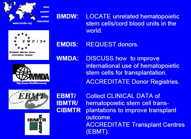
 Copyright © 2001-2014 Dr.
Daniele Focosi. All rights reserved | About
this site | Terms
of use | Legal
notices

 |
 |
 |
 |

|
|
|
|
|||
|
|
|
|||
|
|
|
|
